 Movie
Movie Controller

[English] 日本語
 Yorodumi
- PDB-3pct: Structure of the class C acid phosphatase from Pasteurella multocida -
+ Open data

- Basic information
| Entry | Database: PDB / ID: 3pct | ||||||
|---|---|---|---|---|---|---|---|
| Title | Structure of the class C acid phosphatase from Pasteurella multocida | ||||||
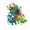
 Components Components | Class C acid phosphatase | ||||||
 Keywords Keywords |  HYDROLASE / outer membrane HYDROLASE / outer membrane | ||||||
| Function / homology |  Function and homology information Function and homology information | ||||||
| Biological species |  Pasteurella multocida (bacteria) Pasteurella multocida (bacteria) | ||||||
| Method | X-RAY DIFFRACTION / SYNCHROTRON / MOLECULAR REPLACEMENT / Resolution: 1.85 Å | ||||||
 Authors Authors | Singh, H. / Malinski, T.J. / Reilly, T.J. / Tanner, J.J. | ||||||
 Citation Citation | Journal: Arch.Biochem.Biophys. / Year: 2011 Title: Crystal structure and immunogenicity of the class C acid phosphatase from Pasteurella multocida. Authors: Singh, H. / Malinski, T.J. / Reilly, T.J. / Henzl, M.T. / Tanner, J.J. | ||||||
| History |
|
- Structure visualization
| Structure viewer | Molecule: MolmilJmol/JSmol |
|---|
- Downloads & links
-Download
| PDBx/mmCIF format | 3pct.cif.gz | 304.7 KB | Display | PDBx/mmCIF format |
|---|---|---|---|---|
| PDB format | pdb3pct.ent.gz | 248.5 KB | Display | PDB format |
| PDBx/mmJSON format | 3pct.json.gz | Tree view | PDBx/mmJSON format | |
| Others |  Other downloads Other downloads |
-Validation report
| Arichive directory | https://data.pdbj.org/pub/pdb/validation_reports/pc/3pctftp://data.pdbj.org/pub/pdb/validation_reports/pc/3pct | HTTPS FTP |
|---|
-Related structure data
| Related structure data |  3et4S S: Starting model for refinement |
|---|---|
| Similar structure data |


-Links
 PDBj
PDBj- Assembly
| Deposited unit | 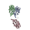
| ||||||||||||
|---|---|---|---|---|---|---|---|---|---|---|---|---|---|
| 1 | 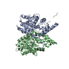
| ||||||||||||
| 2 | 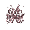
| ||||||||||||
| Unit cell |
| ||||||||||||
| Components on special symmetry positions |
| ||||||||||||
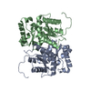
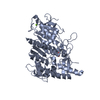
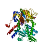
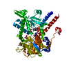
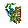
| Details | The asymmetric unit contains a dimer formed by chains A and B. There is a third molecule in the asymmetric unit (chain C), and rotation of it around the crystallographic 2-fold axis (-x, y, -z) followed by translation of (1, 0, 0) generates the CC dimer. |
-Components
| #1: Protein | Mass: 29100.637 Da / Num. of mol.: 3 / Fragment: UNP residues 21-272 Source method: isolated from a genetically manipulated source Source: (gene. exp.) Pasteurella multocida (bacteria) / Gene: acpC / Plasmid: pET20b / Production host: Escherichia coli (E. coli) / Strain (production host): BL21 (DE3) / References: UniProt: B9VWB2, acid phosphatase#2: Water | ChemComp-HOH / | Water Mass: 18.015 Da / Num. of mol.: 573 / Source method: isolated from a natural source / Formula: H2O Mass: 18.015 Da / Num. of mol.: 573 / Source method: isolated from a natural source / Formula: H2O |
|---|
-Experimental details
-Experiment
| Experiment | Method: X-RAY DIFFRACTION / Number of used crystals: 1 |
|---|
- Sample preparation
| Crystal | Density Matthews: 2.18 Å3/Da / Density % sol: 43.55 % |
|---|---|
| Crystal grow | Temperature: 293 K / pH: 7 Details: 20% (w/v) PEG 3350, 0.2 M ammonium citrate, and 10% (v/v) n-propanol, pH 7, temperature 293K |
-Data collection
| Diffraction | Mean temperature: 173 K | ||||||||||||||||||||||||||||||||||||||||||||||||||||||||||||||||||||||||||||||||||||||||
|---|---|---|---|---|---|---|---|---|---|---|---|---|---|---|---|---|---|---|---|---|---|---|---|---|---|---|---|---|---|---|---|---|---|---|---|---|---|---|---|---|---|---|---|---|---|---|---|---|---|---|---|---|---|---|---|---|---|---|---|---|---|---|---|---|---|---|---|---|---|---|---|---|---|---|---|---|---|---|---|---|---|---|---|---|---|---|---|---|---|
| Diffraction source | Source: SYNCHROTRON / Site: ALS  / Beamline: 4.2.2 / Wavelength: 1 Å / Beamline: 4.2.2 / Wavelength: 1 Å | ||||||||||||||||||||||||||||||||||||||||||||||||||||||||||||||||||||||||||||||||||||||||
| Detector | Type: NOIR-1 / Detector: CCD / Date: Aug 1, 2007 | ||||||||||||||||||||||||||||||||||||||||||||||||||||||||||||||||||||||||||||||||||||||||
| Radiation | Protocol: SINGLE WAVELENGTH / Monochromatic (M) / Laue (L): M / Scattering type: x-ray | ||||||||||||||||||||||||||||||||||||||||||||||||||||||||||||||||||||||||||||||||||||||||
| Radiation wavelength | Wavelength: 1 Å / Relative weight: 1 | ||||||||||||||||||||||||||||||||||||||||||||||||||||||||||||||||||||||||||||||||||||||||
| Reflection | Resolution: 1.85→26.616 Å / Num. all: 62294 / Num. obs: 62294 / % possible obs: 97.8 % / Redundancy: 3.5 % / Rmerge(I) obs: 0.094 / Rsym value: 0.094 / Net I/σ(I): 10.4 | ||||||||||||||||||||||||||||||||||||||||||||||||||||||||||||||||||||||||||||||||||||||||
| Reflection shell |
|
- Processing
| Software |
| |||||||||||||||||||||||||||||||||||||||||||||||||||||||||||||||||||||||||||||||||||||||||||||||||||||||||||||||||||||||||||||||||||||||||||||||||||||||||||||||||
|---|---|---|---|---|---|---|---|---|---|---|---|---|---|---|---|---|---|---|---|---|---|---|---|---|---|---|---|---|---|---|---|---|---|---|---|---|---|---|---|---|---|---|---|---|---|---|---|---|---|---|---|---|---|---|---|---|---|---|---|---|---|---|---|---|---|---|---|---|---|---|---|---|---|---|---|---|---|---|---|---|---|---|---|---|---|---|---|---|---|---|---|---|---|---|---|---|---|---|---|---|---|---|---|---|---|---|---|---|---|---|---|---|---|---|---|---|---|---|---|---|---|---|---|---|---|---|---|---|---|---|---|---|---|---|---|---|---|---|---|---|---|---|---|---|---|---|---|---|---|---|---|---|---|---|---|---|---|---|---|---|---|---|
| Refinement | Method to determine structure: MOLECULAR REPLACEMENT Starting model: PDB ENTRY 3ET4 Resolution: 1.85→26.616 Å / Occupancy max: 1 / Occupancy min: 0.02 / SU ML: 0.29 / Cross valid method: THROUGHOUT / σ(F): 1.36 / Stereochemistry target values: ML
| |||||||||||||||||||||||||||||||||||||||||||||||||||||||||||||||||||||||||||||||||||||||||||||||||||||||||||||||||||||||||||||||||||||||||||||||||||||||||||||||||
| Solvent computation | Shrinkage radii: 0.83 Å / VDW probe radii: 1.1 Å / Solvent model: FLAT BULK SOLVENT MODEL / Bsol: 37.529 Å2 / ksol: 0.359 e/Å3 | |||||||||||||||||||||||||||||||||||||||||||||||||||||||||||||||||||||||||||||||||||||||||||||||||||||||||||||||||||||||||||||||||||||||||||||||||||||||||||||||||
| Displacement parameters | Biso max: 106.97 Å2 / Biso mean: 23.5014 Å2 / Biso min: 3.07 Å2
| |||||||||||||||||||||||||||||||||||||||||||||||||||||||||||||||||||||||||||||||||||||||||||||||||||||||||||||||||||||||||||||||||||||||||||||||||||||||||||||||||
| Refinement step | Cycle: LAST / Resolution: 1.85→26.616 Å
| |||||||||||||||||||||||||||||||||||||||||||||||||||||||||||||||||||||||||||||||||||||||||||||||||||||||||||||||||||||||||||||||||||||||||||||||||||||||||||||||||
| Refine LS restraints |
| |||||||||||||||||||||||||||||||||||||||||||||||||||||||||||||||||||||||||||||||||||||||||||||||||||||||||||||||||||||||||||||||||||||||||||||||||||||||||||||||||
| LS refinement shell | Refine-ID: X-RAY DIFFRACTION / Total num. of bins used: 22
| |||||||||||||||||||||||||||||||||||||||||||||||||||||||||||||||||||||||||||||||||||||||||||||||||||||||||||||||||||||||||||||||||||||||||||||||||||||||||||||||||
| Refinement TLS params. | Method: refined / Refine-ID: X-RAY DIFFRACTION
| |||||||||||||||||||||||||||||||||||||||||||||||||||||||||||||||||||||||||||||||||||||||||||||||||||||||||||||||||||||||||||||||||||||||||||||||||||||||||||||||||
| Refinement TLS group |
|
