 Movie
Movie Controller

[English] 日本語
 Yorodumi
- PDB-6k0p: Catalytic domain of GH87 alpha-1,3-glucanase D1045A in complex wi... -
+ Open data

- Basic information
| Entry | Database: PDB / ID: 6k0p | |||||||||
|---|---|---|---|---|---|---|---|---|---|---|
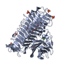
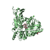
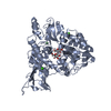
| Title | Catalytic domain of GH87 alpha-1,3-glucanase D1045A in complex with nigerose | |||||||||
 Components Components | Alpha-1,3-glucanase | |||||||||
 Keywords Keywords |  HYDROLASE / GH87 alpha-1 / 3-glucanase / catalytic domain HYDROLASE / GH87 alpha-1 / 3-glucanase / catalytic domain | |||||||||
| Function / homology |  Function and homology information Function and homology information | |||||||||
| Biological species |  Paenibacillus glycanilyticus (bacteria) Paenibacillus glycanilyticus (bacteria) | |||||||||
| Method | X-RAY DIFFRACTION / SYNCHROTRON / MOLECULAR REPLACEMENT / molecular replacement / Resolution: 1.424 Å | |||||||||
 Authors Authors | Itoh, T. / Intuy, R. / Suyotha, W. / Hayashi, J. / Yano, S. / Makabe, K. / Wakayama, M. / Hibi, T. | |||||||||
 Citation Citation | Journal: Febs J. / Year: 2020 Title: Structural insights into substrate recognition and catalysis by glycoside hydrolase family 87 alpha-1,3-glucanase from Paenibacillus glycanilyticus FH11. Authors: Itoh, T. / Intuy, R. / Suyotha, W. / Hayashi, J. / Yano, S. / Makabe, K. / Wakayama, M. / Hibi, T. | |||||||||
| History |
|
- Structure visualization
| Structure viewer | Molecule: MolmilJmol/JSmol |
|---|
- Downloads & links
-Download
| PDBx/mmCIF format | 6k0p.cif.gz | 149.4 KB | Display | PDBx/mmCIF format |
|---|---|---|---|---|
| PDB format | pdb6k0p.ent.gz | 111.1 KB | Display | PDB format |
| PDBx/mmJSON format | 6k0p.json.gz | Tree view | PDBx/mmJSON format | |
| Others |  Other downloads Other downloads |
-Validation report
| Arichive directory | https://data.pdbj.org/pub/pdb/validation_reports/k0/6k0pftp://data.pdbj.org/pub/pdb/validation_reports/k0/6k0p | HTTPS FTP |
|---|
-Related structure data
| Related structure data | 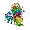 6k0mSC  6k0nC  6k0qC  6k0sC  6k0uC  6k0vC S: Starting model for refinement C: citing same article ( |
|---|---|
| Similar structure data |


-Links
 PDBj
PDBj


- Assembly
| Deposited unit | 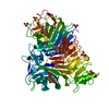
| ||||||||||||
|---|---|---|---|---|---|---|---|---|---|---|---|---|---|
| 1 |
| ||||||||||||
| Unit cell |
| ||||||||||||
| Components on special symmetry positions |
|
-Components
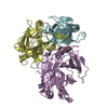
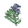
-Protein / Sugars , 2 types, 4 molecules A
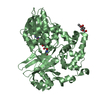
| #1: Protein | Mass: 61935.047 Da / Num. of mol.: 1 / Fragment: Catalytic domain / Mutation: D1045A Source method: isolated from a genetically manipulated source Source: (gene. exp.) Paenibacillus glycanilyticus (bacteria)Gene: agl / Production host: Brevibacillus (bacteria) / References: UniProt: A0A068PS59 |
|---|---|
| #2: Polysaccharide |   , Oligosaccharide / Class: Nutrient / Mass: 342.297 Da / Num. of mol.: 3 , Oligosaccharide / Class: Nutrient / Mass: 342.297 Da / Num. of mol.: 3Source method: isolated from a genetically manipulated source Details: oligosaccharide / References: alpha-nigerose |
-Non-polymers , 4 types, 886 molecules 


| #3: Chemical | ChemComp-SO4 / Sulfate Mass: 96.063 Da / Num. of mol.: 5 / Source method: obtained synthetically / Formula: SO4 Mass: 96.063 Da / Num. of mol.: 5 / Source method: obtained synthetically / Formula: SO4#4: Chemical | ChemComp-CA / |  Mass: 40.078 Da / Num. of mol.: 1 / Source method: obtained synthetically / Formula: Ca Mass: 40.078 Da / Num. of mol.: 1 / Source method: obtained synthetically / Formula: Ca#5: Chemical | Acetic acid Mass: 60.052 Da / Num. of mol.: 2 / Source method: obtained synthetically / Formula: C2H4O2 Mass: 60.052 Da / Num. of mol.: 2 / Source method: obtained synthetically / Formula: C2H4O2#6: Water | ChemComp-HOH / | WaterMass: 18.015 Da / Num. of mol.: 878 / Source method: isolated from a natural source / Formula: H2O |
|---|
-Experimental details
-Experiment
| Experiment | Method: X-RAY DIFFRACTION / Number of used crystals: 1 |
|---|
- Sample preparation
| Crystal | Density Matthews: 2.7 Å3/Da / Density % sol: 54.43 % Description: The entry contains friedel pairs in F_plus/minus columns and I_plus/minus columns |
|---|---|
| Crystal grow | Temperature: 293 K / Method: vapor diffusion, sitting drop / Details: 1.5 M (NH4)2SO4, 25% (v/v) glycerol |
-Data collection
| Diffraction | Mean temperature: 100 K / Serial crystal experiment: N |
|---|---|
| Diffraction source | Source: SYNCHROTRON / Site: SPring-8  / Beamline: BL38B1 / Wavelength: 1 Å / Beamline: BL38B1 / Wavelength: 1 Å |
| Detector | Type: DECTRIS PILATUS3 S 6M / Detector: PIXEL / Date: Oct 31, 2018 |
| Radiation | Protocol: SINGLE WAVELENGTH / Monochromatic (M) / Laue (L): M / Scattering type: x-ray |
| Radiation wavelength | Wavelength: 1 Å / Relative weight: 1 |
| Reflection | Resolution: 1.42→46.855 Å / Num. obs: 125008 / % possible obs: 98.9 % / Redundancy: 4.6 % / Rmerge(I) obs: 0.123 / Net I/σ(I): 9.2 |
| Reflection shell | Resolution: 1.42→1.51 Å / Rmerge(I) obs: 1.254 / Num. unique obs: 38213 |
-Phasing
| Phasing | Method: molecular replacement |
|---|
- Processing
| Software |
| |||||||||||||||||||||||||||||||||||||||||||||||||||||||||||||||||||||||||||||||||||||||||||||||||||||||||||||||||||||||||||||||||||||||||||||||||||||||||||||||||||||||||||||||||||||||||||||||||||||||||||||||||||||||||
|---|---|---|---|---|---|---|---|---|---|---|---|---|---|---|---|---|---|---|---|---|---|---|---|---|---|---|---|---|---|---|---|---|---|---|---|---|---|---|---|---|---|---|---|---|---|---|---|---|---|---|---|---|---|---|---|---|---|---|---|---|---|---|---|---|---|---|---|---|---|---|---|---|---|---|---|---|---|---|---|---|---|---|---|---|---|---|---|---|---|---|---|---|---|---|---|---|---|---|---|---|---|---|---|---|---|---|---|---|---|---|---|---|---|---|---|---|---|---|---|---|---|---|---|---|---|---|---|---|---|---|---|---|---|---|---|---|---|---|---|---|---|---|---|---|---|---|---|---|---|---|---|---|---|---|---|---|---|---|---|---|---|---|---|---|---|---|---|---|---|---|---|---|---|---|---|---|---|---|---|---|---|---|---|---|---|---|---|---|---|---|---|---|---|---|---|---|---|---|---|---|---|---|---|---|---|---|---|---|---|---|---|---|---|---|---|---|---|---|
| Refinement | Method to determine structure: MOLECULAR REPLACEMENT Starting model: 6K0M Resolution: 1.424→36.756 Å / SU ML: 0.13 / Cross valid method: THROUGHOUT / σ(F): 1.34 / Phase error: 16.76 Details: The entry contains friedel pairs in F_plus/minus columns and I_plus/minus columns
| |||||||||||||||||||||||||||||||||||||||||||||||||||||||||||||||||||||||||||||||||||||||||||||||||||||||||||||||||||||||||||||||||||||||||||||||||||||||||||||||||||||||||||||||||||||||||||||||||||||||||||||||||||||||||
| Solvent computation | Shrinkage radii: 0.9 Å / VDW probe radii: 1.11 Å | |||||||||||||||||||||||||||||||||||||||||||||||||||||||||||||||||||||||||||||||||||||||||||||||||||||||||||||||||||||||||||||||||||||||||||||||||||||||||||||||||||||||||||||||||||||||||||||||||||||||||||||||||||||||||
| Displacement parameters | Biso max: 52.11 Å2 / Biso mean: 16.7823 Å2 / Biso min: 4.71 Å2 | |||||||||||||||||||||||||||||||||||||||||||||||||||||||||||||||||||||||||||||||||||||||||||||||||||||||||||||||||||||||||||||||||||||||||||||||||||||||||||||||||||||||||||||||||||||||||||||||||||||||||||||||||||||||||
| Refinement step | Cycle: final / Resolution: 1.424→36.756 Å
| |||||||||||||||||||||||||||||||||||||||||||||||||||||||||||||||||||||||||||||||||||||||||||||||||||||||||||||||||||||||||||||||||||||||||||||||||||||||||||||||||||||||||||||||||||||||||||||||||||||||||||||||||||||||||
| LS refinement shell | Refine-ID: X-RAY DIFFRACTION / Rfactor Rfree error: 0 / Total num. of bins used: 30
|
