 Movie
Movie Controller

[English] 日本語
 Yorodumi
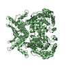
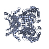
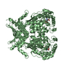
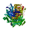
- PDB-3pc4: Full length structure of cystathionine beta-synthase from Drosoph... -
+ Open data

- Basic information
| Entry | Database: PDB / ID: 3pc4 | ||||||
|---|---|---|---|---|---|---|---|
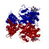
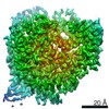
| Title | Full length structure of cystathionine beta-synthase from Drosophila in complex with serine | ||||||
 Components Components | CG1753, isoform A | ||||||
 Keywords Keywords |  LYASE / CBS / Synthase / PLP / Heme / Carbanion LYASE / CBS / Synthase / PLP / Heme / Carbanion | ||||||
| Function / homology |  Function and homology information Function and homology informationCysteine formation from homocysteine / cystathionine beta-synthase / cysteine biosynthetic process via cystathionine / homocysteine metabolic process / cystathionine beta-synthase activity / cysteine synthase activity / L-cysteine desulfhydrase activity / cysteine biosynthetic process from serine / nitrite reductase (NO-forming) activity / response to endoplasmic reticulum stress ...Cysteine formation from homocysteine / cystathionine beta-synthase / cysteine biosynthetic process via cystathionine / homocysteine metabolic process / cystathionine beta-synthase activity / cysteine synthase activity / L-cysteine desulfhydrase activity / cysteine biosynthetic process from serine / nitrite reductase (NO-forming) activity / response to endoplasmic reticulum stress / determination of adult lifespan / metal ion binding / cytoplasmSimilarity search - Function | ||||||
| Biological species |  Drosophila melanogaster (fruit fly) Drosophila melanogaster (fruit fly) | ||||||
| Method | X-RAY DIFFRACTION / SYNCHROTRON / MOLECULAR REPLACEMENT / Resolution: 1.7 Å | ||||||
 Authors Authors | Koutmos, M. / Smith, J.L. | ||||||
 Citation Citation | Journal: Proc.Natl.Acad.Sci.USA / Year: 2010 Title: Structural basis for substrate activation and regulation by cystathionine beta-synthase (CBS) domains in cystathionine {beta}-synthase. Authors: Koutmos, M. / Kabil, O. / Smith, J.L. / Banerjee, R. | ||||||
| History |
|
- Structure visualization
| Structure viewer | Molecule: MolmilJmol/JSmol |
|---|
- Downloads & links
-Download
| PDBx/mmCIF format | 3pc4.cif.gz | 129.5 KB | Display | PDBx/mmCIF format |
|---|---|---|---|---|
| PDB format | pdb3pc4.ent.gz | 95.9 KB | Display | PDB format |
| PDBx/mmJSON format | 3pc4.json.gz | Tree view | PDBx/mmJSON format | |
| Others |  Other downloads Other downloads |
-Validation report
| Arichive directory | https://data.pdbj.org/pub/pdb/validation_reports/pc/3pc4ftp://data.pdbj.org/pub/pdb/validation_reports/pc/3pc4 | HTTPS FTP |
|---|
-Related structure data
| Related structure data | 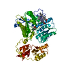 3pc2SC 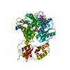 3pc3C S: Starting model for refinement C: citing same article ( |
|---|---|
| Similar structure data |


-Links
 PDBj
PDBj

- Assembly
| Deposited unit | 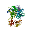
| ||||||||
|---|---|---|---|---|---|---|---|---|---|
| 1 | 
| ||||||||
| Unit cell |
|
-Components
| #1: Protein | Mass: 57469.816 Da / Num. of mol.: 1 Source method: isolated from a genetically manipulated source Source: (gene. exp.) Drosophila melanogaster (fruit fly) / Gene: CG1753, Dmel_CG1753 / Plasmid: pGEX-4T1 / Production host:  Escherichia coli (E. coli) / Strain (production host): BL21 / References: UniProt: Q9VRD9, cystathionine beta-synthase Escherichia coli (E. coli) / Strain (production host): BL21 / References: UniProt: Q9VRD9, cystathionine beta-synthase |
|---|---|
| #2: Chemical | ChemComp-HEM / Heme B  Mass: 616.487 Da / Num. of mol.: 1 / Source method: obtained synthetically / Formula: C34H32FeN4O4 Mass: 616.487 Da / Num. of mol.: 1 / Source method: obtained synthetically / Formula: C34H32FeN4O4 |
| #3: Chemical | ChemComp-KOU / (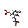  Mass: 334.219 Da / Num. of mol.: 1 / Source method: obtained synthetically / Formula: C11H15N2O8P Mass: 334.219 Da / Num. of mol.: 1 / Source method: obtained synthetically / Formula: C11H15N2O8P |
| #4: Chemical | ChemComp-NA /   Mass: 22.990 Da / Num. of mol.: 1 / Source method: obtained synthetically / Formula: Na Mass: 22.990 Da / Num. of mol.: 1 / Source method: obtained synthetically / Formula: Na |
| #5: Water | ChemComp-HOH / Water Mass: 18.015 Da / Num. of mol.: 598 / Source method: isolated from a natural source / Formula: H2O Mass: 18.015 Da / Num. of mol.: 598 / Source method: isolated from a natural source / Formula: H2O |
| Nonpolymer details | THE UNBIASED ELECTRON DENSITY MAPS IN THE ABSENCE OF THE LIGAND KOU SUGGEST THAT BEST MODELED ...THE UNBIASED ELECTRON DENSITY MAPS IN THE ABSENCE OF THE LIGAND KOU SUGGEST THAT BEST MODELED LIGAND IS THAT OF A CARBANION WITH A PLANAR C-ALPHA. BIOCHEMICA |
-Experimental details
-Experiment
| Experiment | Method: X-RAY DIFFRACTION / Number of used crystals: 1 |
|---|
- Sample preparation
| Crystal | Density Matthews: 2.09 Å3/Da / Density % sol: 41.29 % |
|---|---|
| Crystal grow | Temperature: 298 K / Method: vapor diffusion / pH: 6.5 Details: 23% PEG 3350, 0.2 Li2SO4, 100 mM BIS-TRIS 6.5, vapor diffusion, temperature 298K |
-Data collection
| Diffraction | Mean temperature: 100 K | |||||||||||||||||||||||||||||||||||||||||||||||||||||||||||||||||||||||||||||
|---|---|---|---|---|---|---|---|---|---|---|---|---|---|---|---|---|---|---|---|---|---|---|---|---|---|---|---|---|---|---|---|---|---|---|---|---|---|---|---|---|---|---|---|---|---|---|---|---|---|---|---|---|---|---|---|---|---|---|---|---|---|---|---|---|---|---|---|---|---|---|---|---|---|---|---|---|---|---|
| Diffraction source | Source: SYNCHROTRON / Site: APS  / Beamline: 23-ID-D / Wavelength: 0.9798 Å / Beamline: 23-ID-D / Wavelength: 0.9798 Å | |||||||||||||||||||||||||||||||||||||||||||||||||||||||||||||||||||||||||||||
| Detector | Type: MARMOSAIC 300 mm CCD / Detector: CCD / Date: Dec 17, 2009 Details: K-B pair of biomorph mirrors for vertical and horizontal focusing | |||||||||||||||||||||||||||||||||||||||||||||||||||||||||||||||||||||||||||||
| Radiation | Monochromator: double crystal monochromator / Protocol: SINGLE WAVELENGTH / Monochromatic (M) / Laue (L): M / Scattering type: x-ray | |||||||||||||||||||||||||||||||||||||||||||||||||||||||||||||||||||||||||||||
| Radiation wavelength | Wavelength: 0.9798 Å / Relative weight: 1 | |||||||||||||||||||||||||||||||||||||||||||||||||||||||||||||||||||||||||||||
| Reflection | Resolution: 1.7→50 Å / Num. all: 53048 / Num. obs: 52465 / % possible obs: 98.9 % / Observed criterion σ(F): 0 / Observed criterion σ(I): 0 / Redundancy: 5.4 % / Rmerge(I) obs: 0.063 / Χ2: 0.814 / Net I/σ(I): 11.9 | |||||||||||||||||||||||||||||||||||||||||||||||||||||||||||||||||||||||||||||
| Reflection shell |
|
- Processing
| Software |
| |||||||||||||||||||||||||||||||||||||||||||||||||||||||||||||||||
|---|---|---|---|---|---|---|---|---|---|---|---|---|---|---|---|---|---|---|---|---|---|---|---|---|---|---|---|---|---|---|---|---|---|---|---|---|---|---|---|---|---|---|---|---|---|---|---|---|---|---|---|---|---|---|---|---|---|---|---|---|---|---|---|---|---|---|
| Refinement | Method to determine structure: MOLECULAR REPLACEMENT Starting model: PDB entry 3PC2 Resolution: 1.7→41.21 Å / Cor.coef. Fo:Fc: 0.96 / Cor.coef. Fo:Fc free: 0.951 / WRfactor Rfree: 0.2207 / WRfactor Rwork: 0.1947 / Occupancy max: 1 / Occupancy min: 0.5 / FOM work R set: 0.8714 / SU B: 2.069 / SU ML: 0.069 / SU R Cruickshank DPI: 0.1177 / SU Rfree: 0.1067 / Cross valid method: THROUGHOUT / σ(F): 0 / ESU R Free: 0.107 / Stereochemistry target values: MAXIMUM LIKELIHOOD / Details: U VALUES: REFINED INDIVIDUALLY
| |||||||||||||||||||||||||||||||||||||||||||||||||||||||||||||||||
| Solvent computation | Ion probe radii: 0.8 Å / Shrinkage radii: 0.8 Å / VDW probe radii: 1.4 Å / Solvent model: BABINET MODEL WITH MASK | |||||||||||||||||||||||||||||||||||||||||||||||||||||||||||||||||
| Displacement parameters | Biso max: 62.43 Å2 / Biso mean: 23.1731 Å2 / Biso min: 8.42 Å2
| |||||||||||||||||||||||||||||||||||||||||||||||||||||||||||||||||
| Refinement step | Cycle: LAST / Resolution: 1.7→41.21 Å
| |||||||||||||||||||||||||||||||||||||||||||||||||||||||||||||||||
| Refine LS restraints |
| |||||||||||||||||||||||||||||||||||||||||||||||||||||||||||||||||
| LS refinement shell | Resolution: 1.7→1.747 Å / Total num. of bins used: 20
|
