 Movie
Movie Controller

[English] 日本語
 Yorodumi
- PDB-3pac: Crystal structure of PduT a trimeric bacterial microcompartment p... -
+ Open data

- Basic information
| Entry | Database: PDB / ID: 3pac | ||||||
|---|---|---|---|---|---|---|---|
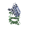
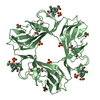
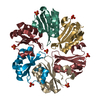
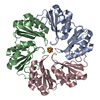
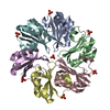
| Title | Crystal structure of PduT a trimeric bacterial microcompartment protein with 4Fe-4S cluster binding site | ||||||
 Components Components | Shell protein | ||||||
 Keywords Keywords |  ELECTRON TRANSPORT / BMC Domain / Shell Protein ELECTRON TRANSPORT / BMC Domain / Shell Protein | ||||||
| Function / homology |  Function and homology information Function and homology informationBacterial microcompartment shell protein PduT / BMC (bacterial microcompartment) domain / BMC domain / Bacterial microcompartment domain / CcmK-like superfamily / BMC / Alpha-Beta Plaits / 2-Layer Sandwich / Alpha BetaSimilarity search - Domain/homology | ||||||
| Biological species |  Citrobacter freundii (bacteria) Citrobacter freundii (bacteria) | ||||||
| Method | X-RAY DIFFRACTION / SYNCHROTRON / SAD / Resolution: 1.86 Å | ||||||
 Authors Authors | Pang, A.H. / Warren, M.J. / Pickersgill, R.W. | ||||||
 Citation Citation | Journal: Acta Crystallogr.,Sect.D / Year: 2011 Title: Structure of PduT, a trimeric bacterial microcompartment protein with a 4Fe-4S cluster-binding site Authors: Pang, A. / Warren, M.J. / Pickersgill, R.W. | ||||||
| History |
|
- Structure visualization
| Structure viewer | Molecule: MolmilJmol/JSmol |
|---|
- Downloads & links
-Download
| PDBx/mmCIF format | 3pac.cif.gz | 46 KB | Display | PDBx/mmCIF format |
|---|---|---|---|---|
| PDB format | pdb3pac.ent.gz | 32.4 KB | Display | PDB format |
| PDBx/mmJSON format | 3pac.json.gz | Tree view | PDBx/mmJSON format | |
| Others |  Other downloads Other downloads |
-Validation report
| Arichive directory | https://data.pdbj.org/pub/pdb/validation_reports/pa/3pacftp://data.pdbj.org/pub/pdb/validation_reports/pa/3pac | HTTPS FTP |
|---|
-Related structure data
| Similar structure data |
|---|

-Links
 PDBj
PDBj

- Assembly
| Deposited unit | 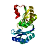
| ||||||||
|---|---|---|---|---|---|---|---|---|---|
| 1 | 
| ||||||||
| Unit cell |
|
-Components
| #1: Protein | Mass: 21226.570 Da / Num. of mol.: 1 Source method: isolated from a genetically manipulated source Source: (gene. exp.) Citrobacter freundii (bacteria) / Gene: pduT / Plasmid: pET14b / Production host: Escherichia coli (E. coli) / Strain (production host): BL21(DE3) / References: UniProt: B1VB78 |
|---|---|
| #2: Water | ChemComp-HOH / Water Mass: 18.015 Da / Num. of mol.: 38 / Source method: isolated from a natural source / Formula: H2O Mass: 18.015 Da / Num. of mol.: 38 / Source method: isolated from a natural source / Formula: H2O |
-Experimental details
-Experiment
| Experiment | Method: X-RAY DIFFRACTION / Number of used crystals: 1 |
|---|
- Sample preparation
| Crystal | Density Matthews: 1.93 Å3/Da / Density % sol: 36.34 % |
|---|---|
| Crystal grow | Temperature: 292 K / Method: vapor diffusion, hanging drop / pH: 8 Details: 0.1M NaCl, 0.1M Bicine, 25% PEG 550, pH 8.0, VAPOR DIFFUSION, HANGING DROP, temperature 292.0K |
-Data collection
| Diffraction |
| ||||||||||||||||||
|---|---|---|---|---|---|---|---|---|---|---|---|---|---|---|---|---|---|---|---|
| Diffraction source |
| ||||||||||||||||||
| Detector |
| ||||||||||||||||||
| Radiation |
| ||||||||||||||||||
| Radiation wavelength |
| ||||||||||||||||||
| Reflection | Resolution: 1.86→33.84 Å / Num. all: 13647 / Num. obs: 13647 / % possible obs: 100 % / Observed criterion σ(F): 0 / Observed criterion σ(I): 0 / Redundancy: 8.2 % / Biso Wilson estimate: 21.2 Å2 / Rmerge(I) obs: 0.035 / Rsym value: 0.095 / Net I/σ(I): 18.5 | ||||||||||||||||||
| Reflection shell | Resolution: 1.86→1.96 Å / Redundancy: 8 % / Rmerge(I) obs: 0.22 / Mean I/σ(I) obs: 3.7 / Num. unique all: 1979 / Rsym value: 0.58 / % possible all: 98 |


- Processing
| Software |
| ||||||||||||||||||||||||||||||||||||||||||||||||||||||||||||||||||||||||||||||||||||||||||||||||||||||||||||||||||||||||||||||||||||||||||||||||||||||||||||||||||||||||||
|---|---|---|---|---|---|---|---|---|---|---|---|---|---|---|---|---|---|---|---|---|---|---|---|---|---|---|---|---|---|---|---|---|---|---|---|---|---|---|---|---|---|---|---|---|---|---|---|---|---|---|---|---|---|---|---|---|---|---|---|---|---|---|---|---|---|---|---|---|---|---|---|---|---|---|---|---|---|---|---|---|---|---|---|---|---|---|---|---|---|---|---|---|---|---|---|---|---|---|---|---|---|---|---|---|---|---|---|---|---|---|---|---|---|---|---|---|---|---|---|---|---|---|---|---|---|---|---|---|---|---|---|---|---|---|---|---|---|---|---|---|---|---|---|---|---|---|---|---|---|---|---|---|---|---|---|---|---|---|---|---|---|---|---|---|---|---|---|---|---|---|---|
| Refinement | Method to determine structure: SAD / Resolution: 1.86→29.71 Å / Cor.coef. Fo:Fc: 0.949 / Cor.coef. Fo:Fc free: 0.921 / SU B: 3.807 / SU ML: 0.115 / Cross valid method: THROUGHOUT / ESU R Free: 0.162 / Stereochemistry target values: MAXIMUM LIKELIHOOD Details: HYDROGENS HAVE BEEN ADDED IN THE RIDING POSITIONS U VALUES: REFINED INDIVIDUALLY
| ||||||||||||||||||||||||||||||||||||||||||||||||||||||||||||||||||||||||||||||||||||||||||||||||||||||||||||||||||||||||||||||||||||||||||||||||||||||||||||||||||||||||||
| Solvent computation | Ion probe radii: 0.8 Å / Shrinkage radii: 0.8 Å / VDW probe radii: 1.4 Å / Solvent model: MASK | ||||||||||||||||||||||||||||||||||||||||||||||||||||||||||||||||||||||||||||||||||||||||||||||||||||||||||||||||||||||||||||||||||||||||||||||||||||||||||||||||||||||||||
| Displacement parameters | Biso mean: 25.825 Å2
| ||||||||||||||||||||||||||||||||||||||||||||||||||||||||||||||||||||||||||||||||||||||||||||||||||||||||||||||||||||||||||||||||||||||||||||||||||||||||||||||||||||||||||
| Refinement step | Cycle: LAST / Resolution: 1.86→29.71 Å
| ||||||||||||||||||||||||||||||||||||||||||||||||||||||||||||||||||||||||||||||||||||||||||||||||||||||||||||||||||||||||||||||||||||||||||||||||||||||||||||||||||||||||||
| Refine LS restraints |
| ||||||||||||||||||||||||||||||||||||||||||||||||||||||||||||||||||||||||||||||||||||||||||||||||||||||||||||||||||||||||||||||||||||||||||||||||||||||||||||||||||||||||||
| LS refinement shell | Resolution: 1.861→1.909 Å / Total num. of bins used: 20
|
