 Movie
Movie Controller

[English] 日本語
 Yorodumi
- PDB-3nwg: The crystal structure of a microcomparments protein from Desulfit... -
+ Open data

- Basic information
| Entry | Database: PDB / ID: 3nwg | ||||||
|---|---|---|---|---|---|---|---|
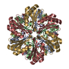
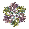
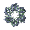
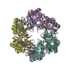
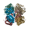
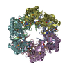
| Title | The crystal structure of a microcomparments protein from Desulfitobacterium hafniense DCB | ||||||
 Components Components | Microcompartments protein Bacterial microcompartment Bacterial microcompartment | ||||||
 Keywords Keywords | STRUCTURAL PROTEIN / Structural Genomics / PSI-2 / Protein Structure Initiative / Midwest Center for Structural Genomics / MCSG / BMC domain | ||||||
| Function / homology |  Function and homology information Function and homology informationBacterial microcompartment shell protein PduT / BMC (bacterial microcompartment) domain / BMC domain / Bacterial microcompartment domain / CcmK-like superfamily / BMC / Alpha-Beta Plaits / 2-Layer Sandwich / Alpha BetaSimilarity search - Domain/homology | ||||||
| Biological species |  Desulfitobacterium hafniense (bacteria) Desulfitobacterium hafniense (bacteria) | ||||||
| Method | X-RAY DIFFRACTION / SYNCHROTRON / SAD / Resolution: 2.7 Å | ||||||
 Authors Authors | Fan, Y. / Volkart, L. / Gu, M. / Axen, S. / Greenleaf, W.B. / Kerfeld, C. / Joachimiak, A. / Midwest Center for Structural Genomics (MCSG) | ||||||
 Citation Citation | Journal: To be Published Title: Structure of a PduT homolog from a novel bacterial microcompartment Authors: Greenleaf, W.B. / Fan, Y. / Axen, S. / Volkart, L. / Gu, M. / Joachimiak, A. / Kerfeld, C. | ||||||
| History |
|
- Structure visualization
| Structure viewer | Molecule: MolmilJmol/JSmol |
|---|
- Downloads & links
-Download
| PDBx/mmCIF format | 3nwg.cif.gz | 70.1 KB | Display | PDBx/mmCIF format |
|---|---|---|---|---|
| PDB format | pdb3nwg.ent.gz | 57.2 KB | Display | PDB format |
| PDBx/mmJSON format | 3nwg.json.gz | Tree view | PDBx/mmJSON format | |
| Others |  Other downloads Other downloads |
-Validation report
| Arichive directory | https://data.pdbj.org/pub/pdb/validation_reports/nw/3nwgftp://data.pdbj.org/pub/pdb/validation_reports/nw/3nwg | HTTPS FTP |
|---|
-Related structure data
| Similar structure data | |
|---|---|
| Other databases |


-Links
 PDBj
PDBj

- Assembly
| Deposited unit | 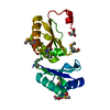
| ||||||||
|---|---|---|---|---|---|---|---|---|---|
| 1 | x 6
| ||||||||
| 2 | 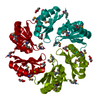
| ||||||||
| Unit cell |
| ||||||||
| Details | Size exclusion chromatography confirms the presence of trimers. Two copies of trimer can potentially stack together and form a hexamer, as found in crystal lattice. |
-Components
| #1: Protein | Bacterial microcompartment Mass: 18957.252 Da / Num. of mol.: 1 Source method: isolated from a genetically manipulated source Source: (gene. exp.) Desulfitobacterium hafniense (bacteria)Strain: DCB / Gene: Dhaf_0360 / Plasmid: pMCSG19 / Production host: Escherichia coli (E. coli) / Strain (production host): pPK1037 / References: UniProt: B8G1Q4 | ||
|---|---|---|---|
| #2: Chemical | Glycerol  Mass: 92.094 Da / Num. of mol.: 3 / Source method: obtained synthetically / Formula: C3H8O3 Mass: 92.094 Da / Num. of mol.: 3 / Source method: obtained synthetically / Formula: C3H8O3#3: Water | ChemComp-HOH / | Water Mass: 18.015 Da / Num. of mol.: 36 / Source method: isolated from a natural source / Formula: H2O Mass: 18.015 Da / Num. of mol.: 36 / Source method: isolated from a natural source / Formula: H2O |
-Experimental details
-Experiment
| Experiment | Method: X-RAY DIFFRACTION / Number of used crystals: 1 |
|---|
- Sample preparation
| Crystal | Density Matthews: 3.4 Å3/Da / Density % sol: 63.86 % |
|---|---|
| Crystal grow | Temperature: 277 K / Method: vapor diffusion, sitting drop / pH: 7.5 Details: 30% (w/v) PEG400, 0.2M magnesium chloride, 0.1M HEPES at pH7.5, VAPOR DIFFUSION, SITTING DROP, temperature 277K |
-Data collection
| Diffraction | Mean temperature: 100 K |
|---|---|
| Diffraction source | Source: SYNCHROTRON / Site: APS  / Beamline: 19-ID / Wavelength: 0.97937 Å / Beamline: 19-ID / Wavelength: 0.97937 Å |
| Detector | Type: ADSC QUANTUM 315 / Detector: CCD / Date: Feb 5, 2009 / Details: mirror |
| Radiation | Monochromator: Si 111 crystal / Protocol: SINGLE WAVELENGTH / Monochromatic (M) / Laue (L): M / Scattering type: x-ray |
| Radiation wavelength | Wavelength: 0.97937 Å / Relative weight: 1 |
| Reflection | Resolution: 2.7→50 Å / Num. all: 7665 / Num. obs: 7665 / % possible obs: 100 % / Observed criterion σ(F): 0 / Observed criterion σ(I): 0 / Redundancy: 20.4 % / Biso Wilson estimate: 54.77 Å2 / Rmerge(I) obs: 0.098 / Net I/σ(I): 53.1 |
| Reflection shell | Resolution: 2.7→2.72 Å / Redundancy: 19.4 % / Rmerge(I) obs: 0.713 / Mean I/σ(I) obs: 4.63 / Num. unique all: 193 / % possible all: 100 |
- Processing
| Software |
| |||||||||||||||||||||||||||||||||||||||||||||||||||||||||||||||||
|---|---|---|---|---|---|---|---|---|---|---|---|---|---|---|---|---|---|---|---|---|---|---|---|---|---|---|---|---|---|---|---|---|---|---|---|---|---|---|---|---|---|---|---|---|---|---|---|---|---|---|---|---|---|---|---|---|---|---|---|---|---|---|---|---|---|---|
| Refinement | Method to determine structure: SAD / Resolution: 2.7→50 Å / Cor.coef. Fo:Fc: 0.93 / Cor.coef. Fo:Fc free: 0.899 / SU B: 25.201 / SU ML: 0.259 / Cross valid method: THROUGHOUT / σ(F): 0 / ESU R: 0.518 / ESU R Free: 0.335 / Stereochemistry target values: MAXIMUM LIKELIHOOD / Details: HYDROGENS HAVE BEEN ADDED IN THE RIDING POSITIONS
| |||||||||||||||||||||||||||||||||||||||||||||||||||||||||||||||||
| Solvent computation | Ion probe radii: 0.8 Å / Shrinkage radii: 0.8 Å / VDW probe radii: 1.4 Å / Solvent model: MASK | |||||||||||||||||||||||||||||||||||||||||||||||||||||||||||||||||
| Displacement parameters | Biso mean: 61.863 Å2
| |||||||||||||||||||||||||||||||||||||||||||||||||||||||||||||||||
| Refinement step | Cycle: LAST / Resolution: 2.7→50 Å
| |||||||||||||||||||||||||||||||||||||||||||||||||||||||||||||||||
| Refine LS restraints |
| |||||||||||||||||||||||||||||||||||||||||||||||||||||||||||||||||
| LS refinement shell | Resolution: 2.702→2.772 Å / Total num. of bins used: 20
| |||||||||||||||||||||||||||||||||||||||||||||||||||||||||||||||||
| Refinement TLS params. | Method: refined / Origin x: 0.6729 Å / Origin y: 40.8598 Å / Origin z: 37.235 Å
| |||||||||||||||||||||||||||||||||||||||||||||||||||||||||||||||||
| Refinement TLS group |
|
