| Entry | Database: PDB / ID: 3p8d
|
|---|
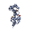
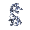
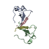
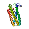
| Title | Crystal structure of the second Tudor domain of human PHF20 (homodimer form) |
|---|
 Components Components | Medulloblastoma antigen MU-MB-50.72 |
|---|
 Keywords Keywords |  PROTEIN BINDING / Tudor domain / LYSINE-METHYLATED P53 BINDING / HISTONE BINDING PROTEIN BINDING / Tudor domain / LYSINE-METHYLATED P53 BINDING / HISTONE BINDING |
|---|
| Function / homology |  Function and homology information Function and homology information
: / NSL complex / Regulation of TP53 Activity through Association with Co-factors / Formation of WDR5-containing histone-modifying complexes / MLL1 complex / histone acetyltransferase complex / Stabilization of p53 / Regulation of TP53 Degradation / chromatin organization / HATs acetylate histones ...: / NSL complex / Regulation of TP53 Activity through Association with Co-factors / Formation of WDR5-containing histone-modifying complexes / MLL1 complex / histone acetyltransferase complex / Stabilization of p53 / Regulation of TP53 Degradation / chromatin organization / HATs acetylate histones / nuclear membrane / regulation of transcription by RNA polymerase II / positive regulation of DNA-templated transcription / DNA binding / nucleoplasm / metal ion binding / cytosolSimilarity search - Function PHD finger protein 20 / PHD finger protein 20, AT hook / PHD finger protein 20, AT-hook / DNA repair protein Crb2, Tudor domain / DNA repair protein Crb2 Tudor domain / PHD finger protein 20-like / PhD finger domain / Tudor domain / Tudor domain / SH3 type barrels. - #140 ...PHD finger protein 20 / PHD finger protein 20, AT hook / PHD finger protein 20, AT-hook / DNA repair protein Crb2, Tudor domain / DNA repair protein Crb2 Tudor domain / PHD finger protein 20-like / PhD finger domain / Tudor domain / Tudor domain / SH3 type barrels. - #140 / Zinc finger C2H2 type domain profile. / Zinc finger, PHD-type, conserved site / Zinc finger C2H2 type domain signature. / Zinc finger PHD-type signature. / Zinc finger C2H2-type / Zinc finger, PHD-type / PHD zinc finger / Zinc finger, FYVE/PHD-type / SH3 type barrels. / Zinc finger, RING/FYVE/PHD-type / Roll / Mainly BetaSimilarity search - Domain/homology |
|---|
| Biological species |  Homo sapiens (human) Homo sapiens (human) |
|---|
| Method | X-RAY DIFFRACTION / SYNCHROTRON / MOLECULAR REPLACEMENT / Resolution: 2 Å |
|---|
 Authors Authors | Cui, G. / Lee, J. / Thompson, J.R. / Botuyan, M.V. / Mer, G. |
|---|
 Citation Citation | Journal: Nat.Struct.Mol.Biol. / Year: 2012
Title: PHF20 is an effector protein of p53 double lysine methylation that stabilizes and activates p53.
Authors: Cui, G. / Park, S. / Badeaux, A.I. / Kim, D. / Lee, J. / Thompson, J.R. / Yan, F. / Kaneko, S. / Yuan, Z. / Botuyan, M.V. / Bedford, M.T. / Cheng, J.Q. / Mer, G. |
|---|
| History | | Deposition | Oct 13, 2010 | Deposition site: RCSB / Processing site: RCSB |
|---|
| Revision 1.0 | Jun 22, 2011 | Provider: repository / Type: Initial release |
|---|
| Revision 1.1 | Jul 13, 2011 | Group: Version format compliance |
|---|
| Revision 1.2 | Aug 15, 2012 | Group: Database references |
|---|
| Revision 1.3 | Sep 26, 2012 | Group: Database references |
|---|
|
|---|
 Movie
Movie Controller

 Yorodumi
 Open data
 Basic information
 Structure visualization
 Downloads & links
 Other downloads


 PDBj
PDBj


 Assembly

 Mass: 18.015 Da / Num. of mol.: 147 / Source method: isolated from a natural source / Formula: H2O
 Sample preparation
 / Beamline: 19-BM / Wavelength: 0.992349 Å
 Processing