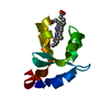
 Movie
Movie Controller


+ Open data

- Basic information
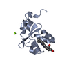
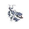
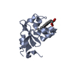
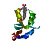
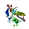
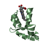
| Entry | Database: PDB / ID: 3ozz | ||||||
|---|---|---|---|---|---|---|---|
| Title | Structure of a cytochrome b5 core-swap mutant | ||||||
 Components Components | Cytochrome b5 | ||||||
 Keywords Keywords | ELECTRON TRANSPORT / HIBRID CYTOCHROME B5 / HEME | ||||||
| Function / homology |  Function and homology information Function and homology informationVitamin C (ascorbate) metabolism / Insertion of tail-anchored proteins into the endoplasmic reticulum membrane / mitochondrial outer membrane / intracellular membrane-bounded organelle / heme binding / endoplasmic reticulum membrane / metal ion bindingSimilarity search - Function | ||||||
| Biological species |  Bos taurus (cattle) Bos taurus (cattle) | ||||||
| Method | X-RAY DIFFRACTION / MOLECULAR REPLACEMENT / Resolution: 1.7 Å | ||||||
 Authors Authors | Terzyan, S. / Parthasarathy, S. / Zhang, X.C. / Benson, D. | ||||||
 Citation Citation | Journal: To be Published Title: Structure of a cytochrome b5 core-swap mutant Authors: Parthasarathy, S. / Terzyan, S. / Zhang, X.C. / Benson, D. | ||||||
| History |
|
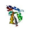
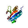
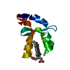
- Structure visualization
| Structure viewer | Molecule: MolmilJmol/JSmol |
|---|
- Downloads & links
-Download
| PDBx/mmCIF format | 3ozz.cif.gz | 36.6 KB | Display | PDBx/mmCIF format |
|---|---|---|---|---|
| PDB format | pdb3ozz.ent.gz | 24 KB | Display | PDB format |
| PDBx/mmJSON format | 3ozz.json.gz | Tree view | PDBx/mmJSON format | |
| Others |  Other downloads Other downloads |
-Validation report
| Arichive directory | https://data.pdbj.org/pub/pdb/validation_reports/oz/3ozzftp://data.pdbj.org/pub/pdb/validation_reports/oz/3ozz | HTTPS FTP |
|---|
-Related structure data
| Related structure data |  1awpS S: Starting model for refinement |
|---|---|
| Similar structure data |


-Links
 PDBj
PDBj
- Assembly
| Deposited unit | 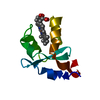
| ||||||||
|---|---|---|---|---|---|---|---|---|---|
| 1 |
| ||||||||
| Unit cell |
| ||||||||
| Components on special symmetry positions |
|
-Components
| #1: Protein | / Cytochrome b5 outer mitochondrial membrane isoform Mass: 9446.489 Da / Num. of mol.: 1 Fragment: N-TERMINAL HEME-BINDING DOMAIN (unp residues 8-89) Source method: isolated from a genetically manipulated source Source: (gene. exp.) Bos taurus (cattle) / Gene: CYB5, CYB5A, Cyb5b, Cyb5m, Omb5 / Plasmid: pET11a / Production host:  Escherichia coli (E. coli) / Strain (production host): Bl21 / References: UniProt: P00171 Escherichia coli (E. coli) / Strain (production host): Bl21 / References: UniProt: P00171 |
|---|---|
| #2: Chemical | ChemComp-HEM / Heme B  Mass: 616.487 Da / Num. of mol.: 1 / Source method: obtained synthetically / Formula: C34H32FeN4O4 Mass: 616.487 Da / Num. of mol.: 1 / Source method: obtained synthetically / Formula: C34H32FeN4O4 |
| #3: Water | ChemComp-HOH / Water Mass: 18.015 Da / Num. of mol.: 142 / Source method: isolated from a natural source / Formula: H2O Mass: 18.015 Da / Num. of mol.: 142 / Source method: isolated from a natural source / Formula: H2O |
-Experimental details
-Experiment
| Experiment | Method: X-RAY DIFFRACTION / Number of used crystals: 1 |
|---|
- Sample preparation
| Crystal | Density Matthews: 2.1 Å3/Da / Density % sol: 41.55 % |
|---|---|
| Crystal grow | Temperature: 277 K / Method: vapor diffusion / pH: 7 Details: 30% PEG8000, 0.2M MgAc, 0.1M Pipes, pH 7.0, VAPOR DIFFUSION, temperature 277K |
-Data collection
| Diffraction | Mean temperature: 80 K |
|---|---|
| Diffraction source | Source: ROTATING ANODE / Type: RIGAKU RUH3R / Wavelength: 1.5418 Å |
| Detector | Type: MAR scanner 345 mm plate / Detector: IMAGE PLATE / Date: Jan 1, 2007 / Details: mirrors |
| Radiation | Monochromator: Osmic Blue optics / Protocol: SINGLE WAVELENGTH / Monochromatic (M) / Laue (L): M / Scattering type: x-ray |
| Radiation wavelength | Wavelength: 1.5418 Å / Relative weight: 1 |
| Reflection | Resolution: 1.7→50 Å / Num. obs: 8707 / % possible obs: 98.4 % / Observed criterion σ(F): -3 / Redundancy: 4 % / Biso Wilson estimate: 17.87 Å2 / Rmerge(I) obs: 0.05 / Net I/σ(I): 24.5 |
| Reflection shell | Resolution: 1.7→1.76 Å / Rmerge(I) obs: 0.152 / Mean I/σ(I) obs: 8.2 / % possible all: 95.3 |
- Processing
| Software |
| ||||||||||||||||||||||||||||||||||||||||||||||||||||||||||||
|---|---|---|---|---|---|---|---|---|---|---|---|---|---|---|---|---|---|---|---|---|---|---|---|---|---|---|---|---|---|---|---|---|---|---|---|---|---|---|---|---|---|---|---|---|---|---|---|---|---|---|---|---|---|---|---|---|---|---|---|---|---|
| Refinement | Method to determine structure: MOLECULAR REPLACEMENT Starting model: PDB ENTRY 1AWP Resolution: 1.7→50 Å / Isotropic thermal model: Anisotropic Overall Temp. factor / σ(F): 0 / Stereochemistry target values: Engh & Huber
| ||||||||||||||||||||||||||||||||||||||||||||||||||||||||||||
| Displacement parameters | Biso mean: 17.72 Å2
| ||||||||||||||||||||||||||||||||||||||||||||||||||||||||||||
| Refinement step | Cycle: LAST / Resolution: 1.7→50 Å
| ||||||||||||||||||||||||||||||||||||||||||||||||||||||||||||
| Refine LS restraints |
|
