 Movie
Movie Controller


+ Open data

- Basic information
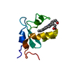
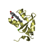
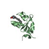
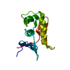
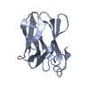
| Entry | Database: PDB / ID: 1icc | ||||||
|---|---|---|---|---|---|---|---|
| Title | RAT OUTER MITOCHONDRIAL MEMBRANE CYTOCHROME B5 | ||||||
 Components Components | CYTOCHROME B5 OUTER MITOCHONDRIAL MEMBRANE ISOFORM | ||||||
 Keywords Keywords |  ELECTRON TRANSPORT / CYTOCHROME / HEME ELECTRON TRANSPORT / CYTOCHROME / HEME | ||||||
| Function / homology |  Function and homology information Function and homology informationSphingolipid de novo biosynthesis / Phase I - Functionalization of compounds / nitric-oxide synthase complex / nitrite reductase (NO-forming) activity / ubiquinol-cytochrome-c reductase activity / enzyme activator activity / nitric oxide biosynthetic process / mitochondrial outer membrane / intracellular membrane-bounded organelle / heme binding / metal ion bindingSimilarity search - Function | ||||||
| Biological species |  Rattus norvegicus (Norway rat) Rattus norvegicus (Norway rat) | ||||||
| Method | X-RAY DIFFRACTION / MOLECULAR REPLACEMENT / Resolution: 2 Å | ||||||
 Authors Authors | Terzyan, S. / Zhang, X. | ||||||
 Citation Citation | Journal: Biochemistry / Year: 2001 Title: Probing the differences between rat liver outer mitochondrial membrane cytochrome b5 and microsomal cytochromes b5. Authors: Altuve, A. / Silchenko, S. / Lee, K.H. / Kuczera, K. / Terzyan, S. / Zhang, X. / Benson, D.R. / Rivera, M. | ||||||
| History |
|
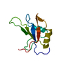
- Structure visualization
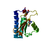
| Structure viewer | Molecule: MolmilJmol/JSmol |
|---|
- Downloads & links
-Download
| PDBx/mmCIF format | 1icc.cif.gz | 92.8 KB | Display | PDBx/mmCIF format |
|---|---|---|---|---|
| PDB format | pdb1icc.ent.gz | 71.1 KB | Display | PDB format |
| PDBx/mmJSON format | 1icc.json.gz | Tree view | PDBx/mmJSON format | |
| Others |  Other downloads Other downloads |
-Validation report
| Arichive directory | https://data.pdbj.org/pub/pdb/validation_reports/ic/1iccftp://data.pdbj.org/pub/pdb/validation_reports/ic/1icc | HTTPS FTP |
|---|
-Related structure data
| Related structure data |  1awpS S: Starting model for refinement |
|---|---|
| Similar structure data |

-Links
 PDBj
PDBj
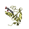
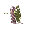
- Assembly
| Deposited unit | 
| ||||||||
|---|---|---|---|---|---|---|---|---|---|
| 1 | 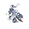
| ||||||||
| 2 | 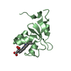
| ||||||||
| 3 | 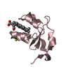
| ||||||||
| 4 | 
| ||||||||
| 5 | 
| ||||||||
| Unit cell |
|
-Components
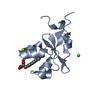
| #1: Protein | Mass: 10006.988 Da / Num. of mol.: 4 / Fragment: WATER SOLUBLE DOMAIN / Mutation: A18S, I32L, L47R Source method: isolated from a genetically manipulated source Source: (gene. exp.) Rattus norvegicus (Norway rat) / Cell: HEPATOCYTE / Organ: LIVER / Plasmid: PET 11A / Species (production host): Escherichia coli / Production host:  Escherichia coli BL21 (bacteria) / Strain (production host): BL21 / References: UniProt: P04166 Escherichia coli BL21 (bacteria) / Strain (production host): BL21 / References: UniProt: P04166#2: Chemical |   Mass: 24.305 Da / Num. of mol.: 3 / Source method: obtained synthetically / Formula: Mg Mass: 24.305 Da / Num. of mol.: 3 / Source method: obtained synthetically / Formula: Mg#3: Chemical | ChemComp-HEM / Heme B  Mass: 616.487 Da / Num. of mol.: 4 / Source method: obtained synthetically / Formula: C34H32FeN4O4 Mass: 616.487 Da / Num. of mol.: 4 / Source method: obtained synthetically / Formula: C34H32FeN4O4#4: Water | ChemComp-HOH / | Water Mass: 18.015 Da / Num. of mol.: 248 / Source method: isolated from a natural source / Formula: H2O Mass: 18.015 Da / Num. of mol.: 248 / Source method: isolated from a natural source / Formula: H2O |
|---|
-Experimental details
-Experiment
| Experiment | Method: X-RAY DIFFRACTION / Number of used crystals: 1 |
|---|
- Sample preparation
| Crystal | Density Matthews: 2.2 Å3/Da / Density % sol: 43 % | ||||||||||||||||||||
|---|---|---|---|---|---|---|---|---|---|---|---|---|---|---|---|---|---|---|---|---|---|
| Crystal grow | Temperature: 278 K / Method: vapor diffusion, hanging drop / pH: 6.5 Details: PEG 8000 Magnesium Acetate PIPES, pH 6.5, VAPOR DIFFUSION, HANGING DROP, temperature 278K | ||||||||||||||||||||
| Crystal grow | *PLUS Temperature: 5 ℃ | ||||||||||||||||||||
| Components of the solutions | *PLUS
|
-Data collection
| Diffraction | Mean temperature: 100 K |
|---|---|
| Diffraction source | Source: ROTATING ANODE / Type: RIGAKU RU300 / Wavelength: 1.5418 Å |
| Detector | Type: MARRESEARCH / Detector: IMAGE PLATE / Date: Feb 10, 2001 / Details: Osmic Blue Optics |
| Radiation | Monochromator: Osmic Mirrors / Protocol: SINGLE WAVELENGTH / Monochromatic (M) / Laue (L): M / Scattering type: x-ray |
| Radiation wavelength | Wavelength: 1.5418 Å / Relative weight: 1 |
| Reflection | Resolution: 2→50 Å / Num. all: 24230 / Num. obs: 23878 / % possible obs: 98.6 % / Observed criterion σ(F): 0 / Observed criterion σ(I): 0 / Redundancy: 3.1 % / Biso Wilson estimate: 29.8 Å2 / Rmerge(I) obs: 0.08 / Net I/σ(I): 12.7 |
| Reflection shell | Resolution: 2→2.07 Å / Redundancy: 3.8 % / Rmerge(I) obs: 0.53 / Mean I/σ(I) obs: 2.5 / Num. unique all: 2324 / % possible all: 98.9 |
| Reflection shell | *PLUS % possible obs: 98.9 % / Num. unique obs: 2324 |
- Processing
| Software |
| |||||||||||||||||||||||||
|---|---|---|---|---|---|---|---|---|---|---|---|---|---|---|---|---|---|---|---|---|---|---|---|---|---|---|
| Refinement | Method to determine structure: MOLECULAR REPLACEMENT Starting model: PDB ENTRY 1AWP Resolution: 2→50 Å / Isotropic thermal model: Anisotropic overall B-Factor / Cross valid method: THROUGHOUT / σ(F): 0 / σ(I): 0 / Stereochemistry target values: Engh and Huber / Details: Used maximum likelihood target using amplitudes
| |||||||||||||||||||||||||
| Solvent computation | Bsol: 53.34 Å2 / ksol: 0.364 e/Å3 | |||||||||||||||||||||||||
| Displacement parameters |
| |||||||||||||||||||||||||
| Refinement step | Cycle: LAST / Resolution: 2→50 Å
| |||||||||||||||||||||||||
| Refine LS restraints |
| |||||||||||||||||||||||||
| LS refinement shell | Resolution: 2→2.02 Å / Total num. of bins used: 29
| |||||||||||||||||||||||||
| Xplor file |
| |||||||||||||||||||||||||
| Software | *PLUS Name: CNS / Classification: refinement | |||||||||||||||||||||||||
| Refinement | *PLUS Highest resolution: 2 Å / Lowest resolution: 50 Å / σ(F): 0 / % reflection Rfree: 6.7 % / Rfactor Rfree: 0.24 | |||||||||||||||||||||||||
| Solvent computation | *PLUS | |||||||||||||||||||||||||
| Displacement parameters | *PLUS | |||||||||||||||||||||||||
| Refine LS restraints | *PLUS Type: c_scangle_it / Dev ideal: 3.2 | |||||||||||||||||||||||||
| LS refinement shell | *PLUS Highest resolution: 2 Å / Rfactor Rfree: 0.326 / % reflection Rfree: 7 % / Rfactor Rwork: 0.278 |
