 Movie
Movie Controller

[English] 日本語
 Yorodumi
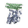
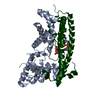
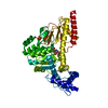
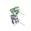
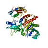
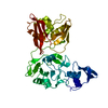
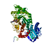
- PDB-3ovu: Crystal Structure of Human Alpha-Haemoglobin Complexed with AHSP ... -
+ Open data

- Basic information
| Entry | Database: PDB / ID: 3ovu | ||||||
|---|---|---|---|---|---|---|---|
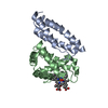
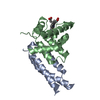
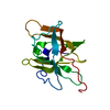
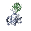
| Title | Crystal Structure of Human Alpha-Haemoglobin Complexed with AHSP and the First NEAT Domain of IsdH from Staphylococcus aureus | ||||||
 Components Components |
| ||||||
 Keywords Keywords | OXYGEN TRANSPORT/PROTEIN BINDING /  haemoglobin / AHSP / NEAT domain / IsdH / protein-protein complex / host-pathogen interaction / OXYGEN TRANSPORT-PROTEIN BINDING complex haemoglobin / AHSP / NEAT domain / IsdH / protein-protein complex / host-pathogen interaction / OXYGEN TRANSPORT-PROTEIN BINDING complex | ||||||
| Function / homology |  Function and homology information Function and homology informationhemoglobin metabolic process / cellular oxidant detoxification / nitric oxide transport / haptoglobin-hemoglobin complex / hemoglobin binding / organic acid binding / hemoglobin complex / hemopoiesis / oxygen transport / Scavenging of heme from plasma ...hemoglobin metabolic process / cellular oxidant detoxification / nitric oxide transport / haptoglobin-hemoglobin complex / hemoglobin binding / organic acid binding / hemoglobin complex / hemopoiesis / oxygen transport / Scavenging of heme from plasma / endocytic vesicle lumen / erythrocyte differentiation / hydrogen peroxide catabolic process / oxygen carrier activity / Heme signaling / carbon dioxide transport / Erythrocytes take up oxygen and release carbon dioxide / response to hydrogen peroxide / Erythrocytes take up carbon dioxide and release oxygen / Cytoprotection by HMOX1 / oxygen binding / unfolded protein binding / protein folding / blood microparticle / protein stabilization / iron ion binding / heme binding / extracellular space / extracellular exosome / extracellular region / membrane / cytosol / cytoplasmSimilarity search - Function | ||||||
| Biological species |  Homo sapiens (human) Homo sapiens (human)  Staphylococcus aureus (bacteria) Staphylococcus aureus (bacteria) | ||||||
| Method | X-RAY DIFFRACTION / SYNCHROTRON / MAD / Resolution: 2.83 Å | ||||||
 Authors Authors | Jacques, D.A. / Krishna Kumar, K. / Caradoc-Davies, T.T. / Langley, D.B. / Mackay, J.P. / Guss, J.M. / Gell, D.A. | ||||||
 Citation Citation | Journal: To be Published Title: A new haem pocket structure in alpha-haemoglobin Authors: Krishna Kumar, K. / Jacques, D.A. / Caradoc-Davies, T.T. / Spirig, T. / Langley, D.B. / Mackay, J.P. / Guss, J.M. / Clubb, R.T. / Gell, D.A. | ||||||
| History |
|
- Structure visualization
| Structure viewer | Molecule: MolmilJmol/JSmol |
|---|
- Downloads & links
-Download
| PDBx/mmCIF format | 3ovu.cif.gz | 163.5 KB | Display | PDBx/mmCIF format |
|---|---|---|---|---|
| PDB format | pdb3ovu.ent.gz | 128 KB | Display | PDB format |
| PDBx/mmJSON format | 3ovu.json.gz | Tree view | PDBx/mmJSON format | |
| Others |  Other downloads Other downloads |
-Validation report
| Arichive directory | https://data.pdbj.org/pub/pdb/validation_reports/ov/3ovuftp://data.pdbj.org/pub/pdb/validation_reports/ov/3ovu | HTTPS FTP |
|---|
-Related structure data
| Related structure data | |
|---|---|
| Similar structure data |


-Links
 PDBj
PDBj


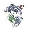
- Assembly
| Deposited unit | 
| ||||||||
|---|---|---|---|---|---|---|---|---|---|
| 1 |
| ||||||||
| Unit cell |
|
-Components
| #1: Protein | Mass: 11721.169 Da / Num. of mol.: 1 Source method: isolated from a genetically manipulated source Source: (gene. exp.) Homo sapiens (human) / Gene: AHSP / Plasmid: pGEX2t / Production host: Escherichia coli (E. coli) / Strain (production host): BL21(DE3) / References: UniProt: Q9NZD4 |
|---|---|
| #2: Protein | Mass: 18904.824 Da / Num. of mol.: 1 / Fragment: first NEAT domain Source method: isolated from a genetically manipulated source Source: (gene. exp.) Staphylococcus aureus (bacteria) / Strain: MSSA476 / Gene: SAS1657 / Plasmid: pET15b / Production host: Escherichia coli (E. coli) / Strain (production host): BL21(DE3) / References: UniProt: Q6G8J7 |
| #3: Protein | / Hemoglobin alpha chain / Alpha-globin Mass: 15150.353 Da / Num. of mol.: 1 / Source method: isolated from a natural source / Details: blood / Source: (natural) Homo sapiens (human) / References: UniProt: P69905 |
| #4: Chemical | ChemComp-HEM / Heme B  Mass: 616.487 Da / Num. of mol.: 1 / Source method: obtained synthetically / Formula: C34H32FeN4O4 Mass: 616.487 Da / Num. of mol.: 1 / Source method: obtained synthetically / Formula: C34H32FeN4O4 |
| #5: Water | ChemComp-HOH / Water Mass: 18.015 Da / Num. of mol.: 3 / Source method: isolated from a natural source / Formula: H2O Mass: 18.015 Da / Num. of mol.: 3 / Source method: isolated from a natural source / Formula: H2O |
-Experimental details
-Experiment
| Experiment | Method: X-RAY DIFFRACTION / Number of used crystals: 1 |
|---|
- Sample preparation
| Crystal | Density Matthews: 2.2 Å3/Da / Density % sol: 44.2 % / Description: The file contains Friedel pair. / Mosaicity: 1.401 ° |
|---|---|
| Crystal grow | Temperature: 293 K / Method: vapor diffusion, hanging drop / pH: 7.5 Details: 0.2M sodium acetate, 20%(w/v) PEG 3350, pH 7.5, VAPOR DIFFUSION, HANGING DROP, temperature 293K |
-Data collection
| Diffraction | Mean temperature: 100 K | |||||||||||||||||||||||||||||||||||||||||||||||||||||||||||||||||||||||||||||||||||||||||||||||||||||||||||||||||||||||||||||||||||||||||||||||||||
|---|---|---|---|---|---|---|---|---|---|---|---|---|---|---|---|---|---|---|---|---|---|---|---|---|---|---|---|---|---|---|---|---|---|---|---|---|---|---|---|---|---|---|---|---|---|---|---|---|---|---|---|---|---|---|---|---|---|---|---|---|---|---|---|---|---|---|---|---|---|---|---|---|---|---|---|---|---|---|---|---|---|---|---|---|---|---|---|---|---|---|---|---|---|---|---|---|---|---|---|---|---|---|---|---|---|---|---|---|---|---|---|---|---|---|---|---|---|---|---|---|---|---|---|---|---|---|---|---|---|---|---|---|---|---|---|---|---|---|---|---|---|---|---|---|---|---|---|---|
| Diffraction source | Source: SYNCHROTRON / Site: Australian Synchrotron  / Beamline: MX2 / Wavelength: 1.74086, 1.73903, 1.67541 / Beamline: MX2 / Wavelength: 1.74086, 1.73903, 1.67541 | |||||||||||||||||||||||||||||||||||||||||||||||||||||||||||||||||||||||||||||||||||||||||||||||||||||||||||||||||||||||||||||||||||||||||||||||||||
| Detector | Type: ADSC QUANTUM 315r / Detector: CCD / Date: May 14, 2010 | |||||||||||||||||||||||||||||||||||||||||||||||||||||||||||||||||||||||||||||||||||||||||||||||||||||||||||||||||||||||||||||||||||||||||||||||||||
| Radiation | Monochromator: Double crystal (Si111) / Protocol: MAD / Monochromatic (M) / Laue (L): M / Scattering type: x-ray | |||||||||||||||||||||||||||||||||||||||||||||||||||||||||||||||||||||||||||||||||||||||||||||||||||||||||||||||||||||||||||||||||||||||||||||||||||
| Radiation wavelength |
| |||||||||||||||||||||||||||||||||||||||||||||||||||||||||||||||||||||||||||||||||||||||||||||||||||||||||||||||||||||||||||||||||||||||||||||||||||
| Reflection | Redundancy: 6.8 % / Av σ(I) over netI: 6.6 / Number: 128528 / Rmerge(I) obs: 0.177 / Χ2: 1.07 / D res high: 2.82 Å / D res low: 50 Å / Num. obs: 18921 / % possible obs: 100 | |||||||||||||||||||||||||||||||||||||||||||||||||||||||||||||||||||||||||||||||||||||||||||||||||||||||||||||||||||||||||||||||||||||||||||||||||||
| Diffraction reflection shell |
| |||||||||||||||||||||||||||||||||||||||||||||||||||||||||||||||||||||||||||||||||||||||||||||||||||||||||||||||||||||||||||||||||||||||||||||||||||
| Reflection | Resolution: 2.83→50 Å / Num. all: 18538 / Num. obs: 18538 / % possible obs: 99.8 % / Observed criterion σ(F): 0 / Observed criterion σ(I): 0 / Redundancy: 3.4 % / Rmerge(I) obs: 0.163 / Χ2: 1.034 / Net I/σ(I): 10.9 | |||||||||||||||||||||||||||||||||||||||||||||||||||||||||||||||||||||||||||||||||||||||||||||||||||||||||||||||||||||||||||||||||||||||||||||||||||
| Reflection shell | Diffraction-ID: 1
|
- Processing
| Software |
| |||||||||||||||||||||||||||||||||||||||||||||||||||||||||||||||||||||||||||||||||||||||||||||||||||||||||||||||||||||||||||||||||||||||||||||||||||||||||||||||||||||||||||||||
|---|---|---|---|---|---|---|---|---|---|---|---|---|---|---|---|---|---|---|---|---|---|---|---|---|---|---|---|---|---|---|---|---|---|---|---|---|---|---|---|---|---|---|---|---|---|---|---|---|---|---|---|---|---|---|---|---|---|---|---|---|---|---|---|---|---|---|---|---|---|---|---|---|---|---|---|---|---|---|---|---|---|---|---|---|---|---|---|---|---|---|---|---|---|---|---|---|---|---|---|---|---|---|---|---|---|---|---|---|---|---|---|---|---|---|---|---|---|---|---|---|---|---|---|---|---|---|---|---|---|---|---|---|---|---|---|---|---|---|---|---|---|---|---|---|---|---|---|---|---|---|---|---|---|---|---|---|---|---|---|---|---|---|---|---|---|---|---|---|---|---|---|---|---|---|---|---|
| Refinement | Method to determine structure: MAD / Resolution: 2.83→49.57 Å / Cor.coef. Fo:Fc: 0.911 / Cor.coef. Fo:Fc free: 0.887 / WRfactor Rfree: 0.3026 / WRfactor Rwork: 0.2785 / Occupancy max: 1 / Occupancy min: 1 / FOM work R set: 0.7802 / SU B: 44.029 / SU ML: 0.395 / SU R Cruickshank DPI: 0.408 / SU Rfree: 0.4532 / Cross valid method: THROUGHOUT / σ(F): 0 / ESU R Free: 0.453 / Stereochemistry target values: MAXIMUM LIKELIHOOD Details: The file contains Friedel pair. HYDROGENS HAVE BEEN ADDED IN THE RIDING POSITIONS U VALUES
| |||||||||||||||||||||||||||||||||||||||||||||||||||||||||||||||||||||||||||||||||||||||||||||||||||||||||||||||||||||||||||||||||||||||||||||||||||||||||||||||||||||||||||||||
| Solvent computation | Ion probe radii: 0.8 Å / Shrinkage radii: 0.8 Å / VDW probe radii: 1.2 Å / Solvent model: MASK | |||||||||||||||||||||||||||||||||||||||||||||||||||||||||||||||||||||||||||||||||||||||||||||||||||||||||||||||||||||||||||||||||||||||||||||||||||||||||||||||||||||||||||||||
| Displacement parameters | Biso max: 69.24 Å2 / Biso mean: 67.188 Å2 / Biso min: 4.36 Å2
| |||||||||||||||||||||||||||||||||||||||||||||||||||||||||||||||||||||||||||||||||||||||||||||||||||||||||||||||||||||||||||||||||||||||||||||||||||||||||||||||||||||||||||||||
| Refinement step | Cycle: LAST / Resolution: 2.83→49.57 Å
| |||||||||||||||||||||||||||||||||||||||||||||||||||||||||||||||||||||||||||||||||||||||||||||||||||||||||||||||||||||||||||||||||||||||||||||||||||||||||||||||||||||||||||||||
| Refine LS restraints |
| |||||||||||||||||||||||||||||||||||||||||||||||||||||||||||||||||||||||||||||||||||||||||||||||||||||||||||||||||||||||||||||||||||||||||||||||||||||||||||||||||||||||||||||||
| LS refinement shell | Resolution: 2.825→2.899 Å / Total num. of bins used: 20
| |||||||||||||||||||||||||||||||||||||||||||||||||||||||||||||||||||||||||||||||||||||||||||||||||||||||||||||||||||||||||||||||||||||||||||||||||||||||||||||||||||||||||||||||
| Refinement TLS params. | Method: refined / Refine-ID: X-RAY DIFFRACTION
| |||||||||||||||||||||||||||||||||||||||||||||||||||||||||||||||||||||||||||||||||||||||||||||||||||||||||||||||||||||||||||||||||||||||||||||||||||||||||||||||||||||||||||||||
| Refinement TLS group |
|
