 Movie
Movie Controller


+ Open data

- Basic information
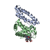
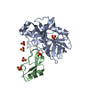
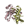
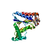
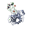
| Entry | Database: PDB / ID: 1z8u | ||||||
|---|---|---|---|---|---|---|---|
| Title | Crystal structure of oxidized alpha hemoglobin bound to AHSP | ||||||
 Components Components |
| ||||||
 Keywords Keywords |  ELECTRON TRANSPORT / alpha haemoglobin / AHSP / oxidation / interaction ELECTRON TRANSPORT / alpha haemoglobin / AHSP / oxidation / interaction | ||||||
| Function / homology |  Function and homology information Function and homology informationhemoglobin metabolic process / cellular oxidant detoxification / nitric oxide transport / haptoglobin-hemoglobin complex / hemoglobin binding / organic acid binding / hemoglobin complex / hemopoiesis / oxygen transport / Scavenging of heme from plasma ...hemoglobin metabolic process / cellular oxidant detoxification / nitric oxide transport / haptoglobin-hemoglobin complex / hemoglobin binding / organic acid binding / hemoglobin complex / hemopoiesis / oxygen transport / Scavenging of heme from plasma / endocytic vesicle lumen / erythrocyte differentiation / hydrogen peroxide catabolic process / oxygen carrier activity / Heme signaling / carbon dioxide transport / Erythrocytes take up oxygen and release carbon dioxide / response to hydrogen peroxide / Erythrocytes take up carbon dioxide and release oxygen / Cytoprotection by HMOX1 / oxygen binding / unfolded protein binding / protein folding / blood microparticle / protein stabilization / iron ion binding / heme binding / extracellular space / extracellular exosome / extracellular region / membrane / cytosol / cytoplasmSimilarity search - Function | ||||||
| Biological species |  Homo sapiens (human) Homo sapiens (human) | ||||||
| Method | X-RAY DIFFRACTION / SYNCHROTRON / MAD / Resolution: 2.4 Å | ||||||
 Authors Authors | Feng, L. / Zhou, S. / Gu, L. / Gell, D.A. / Mackay, J.P. / Weiss, M.J. / Gow, A.J. / Shi, Y. | ||||||
 Citation Citation | Journal: Nature / Year: 2005 Title: Structure of oxidized alpha-haemoglobin bound to AHSP reveals a protective mechanism for haem. Authors: Feng, L. / Zhou, S. / Gu, L. / Gell, D.A. / Mackay, J.P. / Weiss, M.J. / Gow, A.J. / Shi, Y. | ||||||
| History |
|
- Structure visualization
| Structure viewer | Molecule: MolmilJmol/JSmol |
|---|
- Downloads & links
-Download
| PDBx/mmCIF format | 1z8u.cif.gz | 112 KB | Display | PDBx/mmCIF format |
|---|---|---|---|---|
| PDB format | pdb1z8u.ent.gz | 86.2 KB | Display | PDB format |
| PDBx/mmJSON format | 1z8u.json.gz | Tree view | PDBx/mmJSON format | |
| Others |  Other downloads Other downloads |
-Validation report
| Arichive directory | https://data.pdbj.org/pub/pdb/validation_reports/z8/1z8uftp://data.pdbj.org/pub/pdb/validation_reports/z8/1z8u | HTTPS FTP |
|---|
-Related structure data
| Related structure data | |
|---|---|
| Similar structure data |


-Links
 PDBj
PDBj


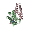
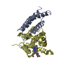
- Assembly
| Deposited unit | 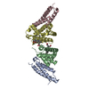
| ||||||||
|---|---|---|---|---|---|---|---|---|---|
| 1 | 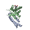
| ||||||||
| 2 | 
| ||||||||
| 3 |
| ||||||||
| Unit cell |
| ||||||||
| Components on special symmetry positions |
|
-Components
| #1: Protein | Mass: 11826.327 Da / Num. of mol.: 2 / Mutation: P30A Source method: isolated from a genetically manipulated source Source: (gene. exp.) Homo sapiens (human) / Gene: AHSP, EDRF, ERAF / Production host:  Escherichia coli (E. coli) / References: UniProt: Q9NZD4 Escherichia coli (E. coli) / References: UniProt: Q9NZD4#2: Protein | Mass: 15281.550 Da / Num. of mol.: 2 Source method: isolated from a genetically manipulated source Source: (gene. exp.) Homo sapiens (human) / Gene: HBA1 / Production host: Escherichia coli (E. coli) / References: UniProt: P69905#3: Chemical | Heme B  Mass: 616.487 Da / Num. of mol.: 2 / Source method: obtained synthetically / Formula: C34H32FeN4O4 Mass: 616.487 Da / Num. of mol.: 2 / Source method: obtained synthetically / Formula: C34H32FeN4O4#4: Water | ChemComp-HOH / | Water Mass: 18.015 Da / Num. of mol.: 388 / Source method: isolated from a natural source / Formula: H2O Mass: 18.015 Da / Num. of mol.: 388 / Source method: isolated from a natural source / Formula: H2O |
|---|
-Experimental details
-Experiment
| Experiment | Method: X-RAY DIFFRACTION / Number of used crystals: 1 |
|---|
- Sample preparation
| Crystal | Density Matthews: 2.4 Å3/Da / Density % sol: 50 % |
|---|---|
| Crystal grow | Temperature: 277 K / Method: vapor diffusion, hanging drop / pH: 6.5 Details: MES, PEG-2000 monomethyl ether, pH 6.5, VAPOR DIFFUSION, HANGING DROP, temperature 277K |
-Data collection
| Diffraction | Mean temperature: 100 K | ||||||||||||
|---|---|---|---|---|---|---|---|---|---|---|---|---|---|
| Diffraction source | Source: SYNCHROTRON / Site: NSLS  / Beamline: X25 / Wavelength: 1.7319, 1.7338, 1.7 / Beamline: X25 / Wavelength: 1.7319, 1.7338, 1.7 | ||||||||||||
| Detector | Type: ADSC QUANTUM 4 / Detector: CCD / Date: Aug 20, 2004 | ||||||||||||
| Radiation | Monochromator: X25 / Protocol: MAD / Monochromatic (M) / Laue (L): M / Scattering type: x-ray | ||||||||||||
| Radiation wavelength |
| ||||||||||||
| Reflection | Resolution: 2.4→99 Å / Num. all: 23106 / Num. obs: 22990 / % possible obs: 99.5 % / Observed criterion σ(F): 0 / Observed criterion σ(I): 0 / Rsym value: 0.058 / Net I/σ(I): 20.9 | ||||||||||||
| Reflection shell | Resolution: 2.4→2.49 Å / Rmerge(I) obs: 0.27 / Mean I/σ(I) obs: 4.5 / Rsym value: 0.27 / % possible all: 99.4 |
- Processing
| Software |
| |||||||||||||||||||||||||
|---|---|---|---|---|---|---|---|---|---|---|---|---|---|---|---|---|---|---|---|---|---|---|---|---|---|---|
| Refinement | Method to determine structure: MAD / Resolution: 2.4→20 Å / σ(F): 0 / σ(I): 0 / Stereochemistry target values: Engh & Huber
| |||||||||||||||||||||||||
| Refinement step | Cycle: LAST / Resolution: 2.4→20 Å
| |||||||||||||||||||||||||
| Refine LS restraints |
|
