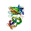
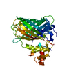
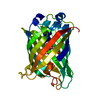
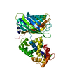
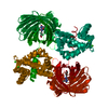
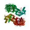
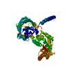
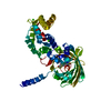
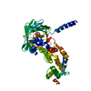
Mass: 46491.883 Da / Num. of mol.: 2 / Mutation: POINT MUTATIONS Source method: isolated from a genetically manipulated source Details: Chimera protein of peptide of M13 (residues 1730-1749, UNP P11799), C-terminal domain of Green fluorescent protein (residues 147-238, UNP P42212), linker, N-terminal of Green fluorescent ...Details: Chimera protein of peptide of M13 (residues 1730-1749, UNP P11799), C-terminal domain of Green fluorescent protein (residues 147-238, UNP P42212), linker, N-terminal of Green fluorescent protein (residues 2-146, UNP P42212) and residues 3-149 of Calmodulin (UNP P0DP23) Source: (gene. exp.) Gallus gallus (chicken), (gene. exp.) Aequorea victoria (jellyfish), (gene. exp.) Homo sapiens (human) Gene: Mylk, GFP, CALM1, CALM, CAM, CAM1 / Plasmid: pET41 / Production host: Escherichia coli (E. coli) / Strain (production host): BL21(DE3) Tuner References: UniProt: P11799, UniProt: P42212, UniProt: P0DP23, myosin-light-chain kinase
Mass: 18.015 Da / Num. of mol.: 77 / Source method: isolated from a natural source / Formula: H2O
Sequence details
UNP RESIDUE 65 SER AND 72 SER OF GREEN FLUORESCENT PROTEIN IN DATABASE UNP P42212 (GFP_AEQVI) ...UNP RESIDUE 65 SER AND 72 SER OF GREEN FLUORESCENT PROTEIN IN DATABASE UNP P42212 (GFP_AEQVI) SHOULD BE GLY. RESIDUE S65G FORM THE CHROMOPHORE CR2 190. RESIDUE S72G (RESIDUE NUMBER 196 IN THE COORDINATES RESPECTIVELY) IS MUTAGENESIS. THESE SEQUENCE WERE BASED ON REFERENCES IN THE DATABASE UNP P42212.
-
Experimental details
-
Experiment
Experiment
Method: X-RAY DIFFRACTION / Number of used crystals: 1
-
Sample preparation
Crystal
Density Matthews: 2.08 Å3/Da / Density % sol: 40.95 %
Crystal grow
Temperature: 293 K / Method: vapor diffusion, hanging drop / pH: 5.5 Details: Reservoir: 100mM Tris HCL (pH 5.5), 100mM (NH4)2SO4, 21% PEG 3350; Protein stock: 7.6 mg/ml Protein, 50mM Tris HCl (pH 7.4), 150mM NaCl; Seed stock solution: 20mM CaCl2, 20% PEG 3350, VAPOR ...Details: Reservoir: 100mM Tris HCL (pH 5.5), 100mM (NH4)2SO4, 21% PEG 3350; Protein stock: 7.6 mg/ml Protein, 50mM Tris HCl (pH 7.4), 150mM NaCl; Seed stock solution: 20mM CaCl2, 20% PEG 3350, VAPOR DIFFUSION, HANGING DROP, temperature 293K
Resolution: 2.6→50.83 Å / Cor.coef. Fo:Fc: 0.904 / Cor.coef. Fo:Fc free: 0.84 / SU B: 24.1 / SU ML: 0.376 / Cross valid method: THROUGHOUT / ESU R Free: 0.45 / Stereochemistry target values: MAXIMUM LIKELIHOOD / Details: HYDROGENS HAVE BEEN ADDED IN THE RIDING POSITIONS
Rfactor
Num. reflection
% reflection
Selection details
Rfree
0.31614
1173
5 %
RANDOM
Rwork
0.24183
-
-
-
obs
0.24546
22288
99.83 %
-
all
-
22326
-
-
Solvent computation
Ion probe radii: 0.8 Å / Shrinkage radii: 0.8 Å / VDW probe radii: 1.2 Å / Solvent model: MASK
In the structure databanks used in Yorodumi, some data are registered as the other names, "COVID-19 virus" and "2019-nCoV". Here are the details of the virus and the list of structure data.
Jan 31, 2019. EMDB accession codes are about to change! (news from PDBe EMDB page)
EMDB accession codes are about to change! (news from PDBe EMDB page)
The allocation of 4 digits for EMDB accession codes will soon come to an end. Whilst these codes will remain in use, new EMDB accession codes will include an additional digit and will expand incrementally as the available range of codes is exhausted. The current 4-digit format prefixed with “EMD-” (i.e. EMD-XXXX) will advance to a 5-digit format (i.e. EMD-XXXXX), and so on. It is currently estimated that the 4-digit codes will be depleted around Spring 2019, at which point the 5-digit format will come into force.
The EM Navigator/Yorodumi systems omit the EMD- prefix.
Related info.:Q: What is EMD? / ID/Accession-code notation in Yorodumi/EM Navigator
Yorodumi is a browser for structure data from EMDB, PDB, SASBDB, etc.
This page is also the successor to EM Navigator detail page, and also detail information page/front-end page for Omokage search.
The word "yorodu" (or yorozu) is an old Japanese word meaning "ten thousand". "mi" (miru) is to see.
Related info.:EMDB / PDB / SASBDB / Comparison of 3 databanks / Yorodumi Search / Aug 31, 2016. New EM Navigator & Yorodumi / Yorodumi Papers / Jmol/JSmol / Function and homology information / Changes in new EM Navigator and Yorodumi
 Movie
Movie Controller


 Open data
 Basic information
 Components
 Myosin light-chain kinase
  Keywords
 Function and homology information


 Authors
 Citation
 Structure visualization
 Downloads & links
 Other downloads
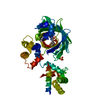


 PDBj
PDBj


 Assembly
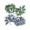


 Mass: 40.078 Da / Num. of mol.: 8 / Source method: obtained synthetically / Formula: Ca
 Mass: 18.015 Da / Num. of mol.: 77 / Source method: isolated from a natural source / Formula: H2O
 Sample preparation
 Processing