 Movie
Movie Controller


+ Open data

- Basic information
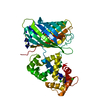
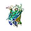
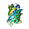
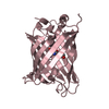
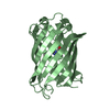
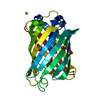
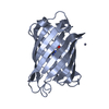
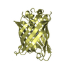
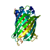
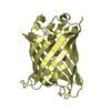
| Entry | Database: PDB / ID: 3evp | ||||||
|---|---|---|---|---|---|---|---|
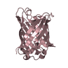
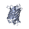
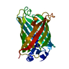
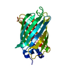
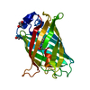
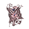
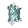
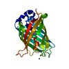
| Title | crystal structure of circular-permutated EGFP | ||||||
 Components Components | Green fluorescent protein,Green fluorescent protein | ||||||
 Keywords Keywords |  SIGNALING PROTEIN / circular-permutated / EGFP / Chromophore / Luminescence / Photoprotein SIGNALING PROTEIN / circular-permutated / EGFP / Chromophore / Luminescence / Photoprotein | ||||||
| Function / homology |  Function and homology information Function and homology information | ||||||
| Biological species |   Aequorea victoria (jellyfish) Aequorea victoria (jellyfish) | ||||||
| Method | X-RAY DIFFRACTION / SYNCHROTRON / MOLECULAR REPLACEMENT / Resolution: 1.453 Å | ||||||
 Authors Authors | Wang, Q. / Shui, B. / Kotlikoff, M.I. / Sondermann, H. | ||||||
 Citation Citation | Journal: Structure / Year: 2008 Title: Structural Basis for Calcium Sensing by GCaMP2. Authors: Wang, Q. / Shui, B. / Kotlikoff, M.I. / Sondermann, H. | ||||||
| History |
|
- Structure visualization
| Structure viewer | Molecule: MolmilJmol/JSmol |
|---|
- Downloads & links
-Download
| PDBx/mmCIF format | 3evp.cif.gz | 122.3 KB | Display | PDBx/mmCIF format |
|---|---|---|---|---|
| PDB format | pdb3evp.ent.gz | 93.7 KB | Display | PDB format |
| PDBx/mmJSON format | 3evp.json.gz | Tree view | PDBx/mmJSON format | |
| Others |  Other downloads Other downloads |
-Validation report
| Arichive directory | https://data.pdbj.org/pub/pdb/validation_reports/ev/3evpftp://data.pdbj.org/pub/pdb/validation_reports/ev/3evp | HTTPS FTP |
|---|
-Related structure data


-Links
 PDBj
PDBj

- Assembly
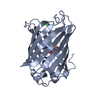
| Deposited unit | 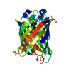
| ||||||||
|---|---|---|---|---|---|---|---|---|---|
| 1 |
| ||||||||
| Unit cell |
|
-Components
| #1: Protein | Mass: 27348.896 Da / Num. of mol.: 1 Source method: isolated from a genetically manipulated source Details: protein contains the M13 fragment of myosin light chain kinase, circular permuted EGFP from Aequorea victoria (Jellyfish), calmodulin from Rattus norvegicus (Rat) Source: (gene. exp.) Aequorea victoria (jellyfish) / Gene: GFP / Plasmid: pET21 / Production host:  Escherichia coli (E. coli) / Strain (production host): BL21(DE3) / References: UniProt: P42212 Escherichia coli (E. coli) / Strain (production host): BL21(DE3) / References: UniProt: P42212 |
|---|---|
| #2: Water | ChemComp-HOH / Water Mass: 18.015 Da / Num. of mol.: 377 / Source method: isolated from a natural source / Formula: H2O Mass: 18.015 Da / Num. of mol.: 377 / Source method: isolated from a natural source / Formula: H2O |
| Sequence details | RESIDUE SER65 HAS BEEN MUTATED TO GLY. RESIDUES GLY65, TYR66, AND GLY67 CONSTITUTE THE CHROMOPHORE ...RESIDUE SER65 HAS BEEN MUTATED TO GLY. RESIDUES GLY65, TYR66, AND GLY67 CONSTITUTE |
-Experimental details
-Experiment
| Experiment | Method: X-RAY DIFFRACTION / Number of used crystals: 5 |
|---|
- Sample preparation
| Crystal | Density Matthews: 2.02 Å3/Da / Density % sol: 39.01 % |
|---|
-Data collection
| Diffraction source | Source: SYNCHROTRON / Site: CHESS  / Beamline: A1 / Beamline: A1 |
|---|---|
| Detector | Type: ADSC QUANTUM 210 / Detector: CCD / Date: Jul 14, 2007 |
| Radiation | Protocol: SINGLE WAVELENGTH / Monochromatic (M) / Laue (L): M / Scattering type: x-ray |
| Radiation wavelength | Relative weight: 1 |
| Reflection | Resolution: 1.45→50 Å / Num. obs: 39659 / Observed criterion σ(I): 9.9 / Redundancy: 7.6 % / Biso Wilson estimate: 13.4 Å2 / Rsym value: 0.042 |
- Processing
| Software |
| ||||||||||||||||||||||||||||||||||||||||||||||||||||||||||||||||||||||||||||||||||||||||||||||||||||||||||||||||||||||||||||||||||||||||||||||||||||||||||||||||||||||||
|---|---|---|---|---|---|---|---|---|---|---|---|---|---|---|---|---|---|---|---|---|---|---|---|---|---|---|---|---|---|---|---|---|---|---|---|---|---|---|---|---|---|---|---|---|---|---|---|---|---|---|---|---|---|---|---|---|---|---|---|---|---|---|---|---|---|---|---|---|---|---|---|---|---|---|---|---|---|---|---|---|---|---|---|---|---|---|---|---|---|---|---|---|---|---|---|---|---|---|---|---|---|---|---|---|---|---|---|---|---|---|---|---|---|---|---|---|---|---|---|---|---|---|---|---|---|---|---|---|---|---|---|---|---|---|---|---|---|---|---|---|---|---|---|---|---|---|---|---|---|---|---|---|---|---|---|---|---|---|---|---|---|---|---|---|---|---|---|---|---|
| Refinement | Method to determine structure: MOLECULAR REPLACEMENT / Resolution: 1.453→41.138 Å / SU ML: 0.15 / σ(F): 0.21 / Phase error: 14.32 / Stereochemistry target values: ML
| ||||||||||||||||||||||||||||||||||||||||||||||||||||||||||||||||||||||||||||||||||||||||||||||||||||||||||||||||||||||||||||||||||||||||||||||||||||||||||||||||||||||||
| Solvent computation | Shrinkage radii: 0.9 Å / VDW probe radii: 1.11 Å / Solvent model: FLAT BULK SOLVENT MODEL / Bsol: 68.075 Å2 / ksol: 0.396 e/Å3 | ||||||||||||||||||||||||||||||||||||||||||||||||||||||||||||||||||||||||||||||||||||||||||||||||||||||||||||||||||||||||||||||||||||||||||||||||||||||||||||||||||||||||
| Refinement step | Cycle: LAST / Resolution: 1.453→41.138 Å
| ||||||||||||||||||||||||||||||||||||||||||||||||||||||||||||||||||||||||||||||||||||||||||||||||||||||||||||||||||||||||||||||||||||||||||||||||||||||||||||||||||||||||
| Refine LS restraints |
| ||||||||||||||||||||||||||||||||||||||||||||||||||||||||||||||||||||||||||||||||||||||||||||||||||||||||||||||||||||||||||||||||||||||||||||||||||||||||||||||||||||||||
| LS refinement shell |
|
