 Movie
Movie Controller

[English] 日本語
 Yorodumi
- PDB-3o0t: Crystal structure of human phosphoglycerate mutase family member ... -
+ Open data

- Basic information
| Entry | Database: PDB / ID: 3o0t | ||||||
|---|---|---|---|---|---|---|---|
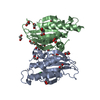
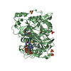
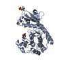
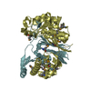
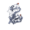
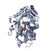
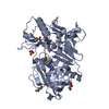
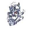
| Title | Crystal structure of human phosphoglycerate mutase family member 5 (PGAM5) in complex with phosphate | ||||||
 Components Components | Serine/threonine-protein phosphatase PGAM5, mitochondrial | ||||||
 Keywords Keywords |  HYDROLASE / Structural Genomics / Structural Genomics Consortium / SGC / Phosphoglycerate mutase family member 5 / PGAM5 / BXLBv68 / MGC5352 protein / Serine/Threonine phosphatase / mitochondrial protein HYDROLASE / Structural Genomics / Structural Genomics Consortium / SGC / Phosphoglycerate mutase family member 5 / PGAM5 / BXLBv68 / MGC5352 protein / Serine/Threonine phosphatase / mitochondrial protein | ||||||
| Function / homology |  Function and homology information Function and homology informationnegative regulation of cold-induced thermogenesis / myosin phosphatase activity / positive regulation of mitochondrial fission / Receptor Mediated Mitophagy / protein serine/threonine phosphatase activity / protein-serine/threonine phosphatase / phosphatase activity / necroptotic process / GTPase activator activity / macroautophagy ...negative regulation of cold-induced thermogenesis / myosin phosphatase activity / positive regulation of mitochondrial fission / Receptor Mediated Mitophagy / protein serine/threonine phosphatase activity / protein-serine/threonine phosphatase / phosphatase activity / necroptotic process / GTPase activator activity / macroautophagy / mitochondrial outer membrane / mitochondrial inner membrane / protein-containing complex binding / mitochondrionSimilarity search - Function | ||||||
| Biological species |  Homo sapiens (human) Homo sapiens (human) | ||||||
| Method | X-RAY DIFFRACTION / MOLECULAR REPLACEMENT / Resolution: 1.9 Å | ||||||
 Authors Authors | Chaikuad, A. / Alfano, I. / Picaud, S. / Filippakopoulos, P. / Barr, A. / von Delft, F. / Arrowsmith, C.H. / Edwards, A.M. / Weigelt, J. / Bountra, C. ...Chaikuad, A. / Alfano, I. / Picaud, S. / Filippakopoulos, P. / Barr, A. / von Delft, F. / Arrowsmith, C.H. / Edwards, A.M. / Weigelt, J. / Bountra, C. / Takeda, K. / Ichijo, H. / Knapp, S. / Structural Genomics Consortium (SGC) | ||||||
 Citation Citation | Journal: Structure / Year: 2017 Title: Structures of PGAM5 Provide Insight into Active Site Plasticity and Multimeric Assembly. Authors: Chaikuad, A. / Filippakopoulos, P. / Marcsisin, S.R. / Picaud, S. / Schroder, M. / Sekine, S. / Ichijo, H. / Engen, J.R. / Takeda, K. / Knapp, S. | ||||||
| History |
|
- Structure visualization
| Structure viewer | Molecule: MolmilJmol/JSmol |
|---|
- Downloads & links
-Download
| PDBx/mmCIF format | 3o0t.cif.gz | 173.2 KB | Display | PDBx/mmCIF format |
|---|---|---|---|---|
| PDB format | pdb3o0t.ent.gz | 138.6 KB | Display | PDB format |
| PDBx/mmJSON format | 3o0t.json.gz | Tree view | PDBx/mmJSON format | |
| Others |  Other downloads Other downloads |
-Validation report
| Arichive directory | https://data.pdbj.org/pub/pdb/validation_reports/o0/3o0tftp://data.pdbj.org/pub/pdb/validation_reports/o0/3o0t | HTTPS FTP |
|---|
-Related structure data


-Links
 PDBj
PDBj


- Assembly
| Deposited unit | 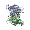
| ||||||||
|---|---|---|---|---|---|---|---|---|---|
| 1 |
| ||||||||
| 2 | 
| ||||||||
| Unit cell |
|
-Components
| #1: Protein | Mass: 23003.281 Da / Num. of mol.: 2 / Fragment: UNP residues 90-289 Source method: isolated from a genetically manipulated source Source: (gene. exp.) Homo sapiens (human) / Gene: PGAM5 / Plasmid: pNIC28-Bsa4 / Production host:  Escherichia coli (E. coli) / Strain (production host): BL21(DE3)-R3 Escherichia coli (E. coli) / Strain (production host): BL21(DE3)-R3References: UniProt: Q96HS1, protein-serine/threonine phosphatase #2: Chemical | ChemComp-PO4 / Phosphate  Mass: 94.971 Da / Num. of mol.: 4 / Source method: obtained synthetically / Formula: PO4 Mass: 94.971 Da / Num. of mol.: 4 / Source method: obtained synthetically / Formula: PO4#3: Chemical | ChemComp-EDO / Ethylene glycol  Mass: 62.068 Da / Num. of mol.: 11 / Source method: obtained synthetically / Formula: C2H6O2 Mass: 62.068 Da / Num. of mol.: 11 / Source method: obtained synthetically / Formula: C2H6O2#4: Water | ChemComp-HOH / | Water Mass: 18.015 Da / Num. of mol.: 178 / Source method: isolated from a natural source / Formula: H2O Mass: 18.015 Da / Num. of mol.: 178 / Source method: isolated from a natural source / Formula: H2O |
|---|
-Experimental details
-Experiment
| Experiment | Method: X-RAY DIFFRACTION / Number of used crystals: 1 |
|---|
- Sample preparation
| Crystal | Density Matthews: 2.29 Å3/Da / Density % sol: 46.31 % |
|---|---|
| Crystal grow | Temperature: 293 K / Method: vapor diffusion, sitting drop / pH: 7.5 Details: 15% PEG-smear (PEG3350 & PEG MME5K), 0.1M HEPES pH 7.5, VAPOR DIFFUSION, SITTING DROP, temperature 293K |
-Data collection
| Diffraction | Mean temperature: 100 K |
|---|---|
| Diffraction source | Source: ROTATING ANODE / Type: RIGAKU FR-E SUPERBRIGHT / Wavelength: 1.542 Å |
| Detector | Type: RIGAKU RAXIS IV / Detector: IMAGE PLATE / Date: Dec 13, 2009 |
| Radiation | Monochromator: Flat graphite crystal / Protocol: SINGLE WAVELENGTH / Monochromatic (M) / Laue (L): M / Scattering type: x-ray |
| Radiation wavelength | Wavelength: 1.542 Å / Relative weight: 1 |
| Reflection | Resolution: 1.9→36.54 Å / Num. all: 34056 / Num. obs: 33999 / % possible obs: 100 % / Observed criterion σ(F): 0 / Observed criterion σ(I): 0 / Redundancy: 4.7 % / Biso Wilson estimate: 34.7 Å2 / Rmerge(I) obs: 0.049 / Net I/σ(I): 15.8 |
| Reflection shell | Resolution: 1.9→2 Å / Redundancy: 4.6 % / Rmerge(I) obs: 0.669 / Mean I/σ(I) obs: 2.2 / Num. unique all: 4896 / % possible all: 100 |
- Processing
| Software |
| |||||||||||||||||||||||||||||||||||||||||||||||||||||||||||||||||||||||||||||||||||||||||||||||||||||||||||||||||||||||||||||||||||||||||||||||||||||||||||||||||||||||||||||||
|---|---|---|---|---|---|---|---|---|---|---|---|---|---|---|---|---|---|---|---|---|---|---|---|---|---|---|---|---|---|---|---|---|---|---|---|---|---|---|---|---|---|---|---|---|---|---|---|---|---|---|---|---|---|---|---|---|---|---|---|---|---|---|---|---|---|---|---|---|---|---|---|---|---|---|---|---|---|---|---|---|---|---|---|---|---|---|---|---|---|---|---|---|---|---|---|---|---|---|---|---|---|---|---|---|---|---|---|---|---|---|---|---|---|---|---|---|---|---|---|---|---|---|---|---|---|---|---|---|---|---|---|---|---|---|---|---|---|---|---|---|---|---|---|---|---|---|---|---|---|---|---|---|---|---|---|---|---|---|---|---|---|---|---|---|---|---|---|---|---|---|---|---|---|---|---|---|
| Refinement | Method to determine structure: MOLECULAR REPLACEMENT / Resolution: 1.9→36.54 Å / Cor.coef. Fo:Fc: 0.968 / Cor.coef. Fo:Fc free: 0.945 / SU B: 6.974 / SU ML: 0.102 / Cross valid method: THROUGHOUT / σ(F): 0 / ESU R Free: 0.142 / Stereochemistry target values: MAXIMUM LIKELIHOOD Details: HYDROGENS HAVE BEEN ADDED IN THE RIDING POSITIONS BUT NOT OUTPUT TO COORDINATE FILE
| |||||||||||||||||||||||||||||||||||||||||||||||||||||||||||||||||||||||||||||||||||||||||||||||||||||||||||||||||||||||||||||||||||||||||||||||||||||||||||||||||||||||||||||||
| Solvent computation | Ion probe radii: 0.8 Å / Shrinkage radii: 0.8 Å / VDW probe radii: 1.2 Å / Solvent model: MASK | |||||||||||||||||||||||||||||||||||||||||||||||||||||||||||||||||||||||||||||||||||||||||||||||||||||||||||||||||||||||||||||||||||||||||||||||||||||||||||||||||||||||||||||||
| Displacement parameters | Biso mean: 38.081 Å2
| |||||||||||||||||||||||||||||||||||||||||||||||||||||||||||||||||||||||||||||||||||||||||||||||||||||||||||||||||||||||||||||||||||||||||||||||||||||||||||||||||||||||||||||||
| Refine analyze | Luzzati coordinate error obs: 0.246 Å | |||||||||||||||||||||||||||||||||||||||||||||||||||||||||||||||||||||||||||||||||||||||||||||||||||||||||||||||||||||||||||||||||||||||||||||||||||||||||||||||||||||||||||||||
| Refinement step | Cycle: LAST / Resolution: 1.9→36.54 Å
| |||||||||||||||||||||||||||||||||||||||||||||||||||||||||||||||||||||||||||||||||||||||||||||||||||||||||||||||||||||||||||||||||||||||||||||||||||||||||||||||||||||||||||||||
| Refine LS restraints |
| |||||||||||||||||||||||||||||||||||||||||||||||||||||||||||||||||||||||||||||||||||||||||||||||||||||||||||||||||||||||||||||||||||||||||||||||||||||||||||||||||||||||||||||||
| LS refinement shell | Resolution: 1.9→1.949 Å / Total num. of bins used: 20
| |||||||||||||||||||||||||||||||||||||||||||||||||||||||||||||||||||||||||||||||||||||||||||||||||||||||||||||||||||||||||||||||||||||||||||||||||||||||||||||||||||||||||||||||
| Refinement TLS params. | Method: refined / Refine-ID: X-RAY DIFFRACTION
| |||||||||||||||||||||||||||||||||||||||||||||||||||||||||||||||||||||||||||||||||||||||||||||||||||||||||||||||||||||||||||||||||||||||||||||||||||||||||||||||||||||||||||||||
| Refinement TLS group |
|
