 Movie
Movie Controller


+ Open data

- Basic information
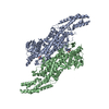
| Entry | Database: PDB / ID: 3nz4 | |||||||||
|---|---|---|---|---|---|---|---|---|---|---|
| Title | Crystal Structure of a Taxus Phenylalanine Aminomutase | |||||||||
 Components Components | Phenylalanine ammonia-lyase | |||||||||
 Keywords Keywords | LYASE / Aminomutase / Taxol biosynthetic pathway / MIO / phenylalanine | |||||||||
| Function / homology |  Function and homology information Function and homology informationphenylalanine aminomutase (L-beta-phenylalanine forming) / intramolecular aminotransferase activity / paclitaxel biosynthetic process / phenylalanine ammonia-lyase / L-phenylalanine metabolic process / cinnamic acid biosynthetic process / alkaloid biosynthetic process / phenylalanine ammonia-lyase activity / L-phenylalanine catabolic process / protein homotetramerization / cytoplasmSimilarity search - Function | |||||||||
| Biological species |  Taxus canadensis (Canada yew) Taxus canadensis (Canada yew) | |||||||||
| Method | X-RAY DIFFRACTION / SYNCHROTRON / MOLECULAR REPLACEMENT / Resolution: 2.38 Å | |||||||||
 Authors Authors | Feng, L. / Geiger, J.H. | |||||||||
 Citation Citation | Journal: Biochemistry / Year: 2011 Title: Mechanistic, mutational, and structural evaluation of a taxus phenylalanine aminomutase. Authors: Feng, L. / Wanninayake, U. / Strom, S. / Geiger, J. / Walker, K.D. | |||||||||
| History |
|
- Structure visualization
| Structure viewer | Molecule: MolmilJmol/JSmol |
|---|
- Downloads & links
-Download
| PDBx/mmCIF format | 3nz4.cif.gz | 509.9 KB | Display | PDBx/mmCIF format |
|---|---|---|---|---|
| PDB format | pdb3nz4.ent.gz | 422.8 KB | Display | PDB format |
| PDBx/mmJSON format | 3nz4.json.gz | Tree view | PDBx/mmJSON format | |
| Others |  Other downloads Other downloads |
-Validation report
| Arichive directory | https://data.pdbj.org/pub/pdb/validation_reports/nz/3nz4ftp://data.pdbj.org/pub/pdb/validation_reports/nz/3nz4 | HTTPS FTP |
|---|
-Related structure data
| Similar structure data |
|---|


-Links
 PDBj
PDBj

- Assembly
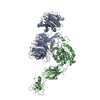
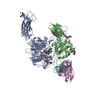
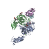
| Deposited unit | 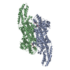
| |||||||||
|---|---|---|---|---|---|---|---|---|---|---|
| 1 |
| |||||||||
| 2 | 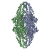
| |||||||||
| Unit cell |
| |||||||||
| Components on special symmetry positions |
|
-Components
| #1: Protein | Mass: 76594.492 Da / Num. of mol.: 2 Source method: isolated from a genetically manipulated source Source: (gene. exp.) Taxus canadensis (Canada yew) / Production host:  Escherichia coli (E. coli) / References: UniProt: Q6GZ04, phenylalanine ammonia-lyase Escherichia coli (E. coli) / References: UniProt: Q6GZ04, phenylalanine ammonia-lyase#2: Chemical | Cinnamic acid  Mass: 148.159 Da / Num. of mol.: 2 / Source method: obtained synthetically / Formula: C9H8O2 Mass: 148.159 Da / Num. of mol.: 2 / Source method: obtained synthetically / Formula: C9H8O2#3: Water | ChemComp-HOH / | Water Mass: 18.015 Da / Num. of mol.: 276 / Source method: isolated from a natural source / Formula: H2O Mass: 18.015 Da / Num. of mol.: 276 / Source method: isolated from a natural source / Formula: H2O |
|---|
-Experimental details
-Experiment
| Experiment | Method: X-RAY DIFFRACTION / Number of used crystals: 1 |
|---|
- Sample preparation
| Crystal | Density Matthews: 2.34 Å3/Da / Density % sol: 47.51 % |
|---|---|
| Crystal grow | Temperature: 298 K / Method: vapor diffusion, sitting drop / pH: 7 Details: 0.1M HEPES, pH 7.0, 1.0M LiCl, 15% PEG 6000, VAPOR DIFFUSION, SITTING DROP, temperature 298.0K |
-Data collection
| Diffraction | Mean temperature: 100 K |
|---|---|
| Diffraction source | Source: SYNCHROTRON / Site: APS  / Beamline: 21-ID-D / Wavelength: 1.0782 Å / Beamline: 21-ID-D / Wavelength: 1.0782 Å |
| Detector | Type: MARMOSAIC 300 mm CCD / Detector: CCD / Date: Apr 7, 2010 |
| Radiation | Protocol: SINGLE WAVELENGTH / Monochromatic (M) / Laue (L): M / Scattering type: x-ray |
| Radiation wavelength | Wavelength: 1.0782 Å / Relative weight: 1 |
| Reflection | Resolution: 2.38→103.81 Å / Num. all: 55249 / Num. obs: 49662 / % possible obs: 96.8 % / Observed criterion σ(F): 2 / Observed criterion σ(I): 1 |
| Reflection shell | Resolution: 2.38→2.42 Å / % possible all: 79.6 |
- Processing
| Software |
| |||||||||||||||||||||||||||||||||||||||||||||||||||||||||||||||||||||||||||||||||||||||||||||||||||||||||||||||||||||||||||||
|---|---|---|---|---|---|---|---|---|---|---|---|---|---|---|---|---|---|---|---|---|---|---|---|---|---|---|---|---|---|---|---|---|---|---|---|---|---|---|---|---|---|---|---|---|---|---|---|---|---|---|---|---|---|---|---|---|---|---|---|---|---|---|---|---|---|---|---|---|---|---|---|---|---|---|---|---|---|---|---|---|---|---|---|---|---|---|---|---|---|---|---|---|---|---|---|---|---|---|---|---|---|---|---|---|---|---|---|---|---|---|---|---|---|---|---|---|---|---|---|---|---|---|---|---|---|---|
| Refinement | Method to determine structure: MOLECULAR REPLACEMENT / Resolution: 2.38→103.81 Å / Cor.coef. Fo:Fc: 0.955 / Cor.coef. Fo:Fc free: 0.926 / SU B: 17.906 / SU ML: 0.187 / Cross valid method: THROUGHOUT / σ(F): 5 / ESU R Free: 0.274 / Stereochemistry target values: MAXIMUM LIKELIHOOD / Details: HYDROGENS HAVE BEEN ADDED IN THE RIDING POSITIONS
| |||||||||||||||||||||||||||||||||||||||||||||||||||||||||||||||||||||||||||||||||||||||||||||||||||||||||||||||||||||||||||||
| Solvent computation | Ion probe radii: 0.8 Å / Shrinkage radii: 0.8 Å / VDW probe radii: 1.4 Å / Solvent model: BABINET MODEL WITH MASK | |||||||||||||||||||||||||||||||||||||||||||||||||||||||||||||||||||||||||||||||||||||||||||||||||||||||||||||||||||||||||||||
| Displacement parameters | Biso mean: 47.117 Å2
| |||||||||||||||||||||||||||||||||||||||||||||||||||||||||||||||||||||||||||||||||||||||||||||||||||||||||||||||||||||||||||||
| Refinement step | Cycle: LAST / Resolution: 2.38→103.81 Å
| |||||||||||||||||||||||||||||||||||||||||||||||||||||||||||||||||||||||||||||||||||||||||||||||||||||||||||||||||||||||||||||
| Refine LS restraints |
| |||||||||||||||||||||||||||||||||||||||||||||||||||||||||||||||||||||||||||||||||||||||||||||||||||||||||||||||||||||||||||||
| LS refinement shell | Resolution: 2.38→2.442 Å / Total num. of bins used: 20
| |||||||||||||||||||||||||||||||||||||||||||||||||||||||||||||||||||||||||||||||||||||||||||||||||||||||||||||||||||||||||||||
| Refinement TLS params. | Method: refined / Refine-ID: X-RAY DIFFRACTION
| |||||||||||||||||||||||||||||||||||||||||||||||||||||||||||||||||||||||||||||||||||||||||||||||||||||||||||||||||||||||||||||
| Refinement TLS group |
|
