Mass: 18.015 Da / Num. of mol.: 1071 / Source method: isolated from a natural source / Formula: H2O
Compound details
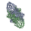
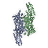
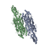
ENZYME OF PLANT METABOLISM CATALYZING THE FIRST REACTION IN THE BIOSYNTHESIS FROM L-PHENYLALANINE. ...ENZYME OF PLANT METABOLISM CATALYZING THE FIRST REACTION IN THE BIOSYNTHESIS FROM L-PHENYLALANINE. CONTAINS AN ACTIVE SITE 4-METHYLIDENE-IMIDAZOLE-5-ONE (MIO), WHICH IS FORMED AUTOCATALYTICALLY BY CYCLIZATION AND DEHYDRATION OF RESIDUES ALA-SER-GLY.
-
Experimental details
-
Experiment
Experiment
Method: X-RAY DIFFRACTION / Number of used crystals: 1
 Movie
Movie Controller


 Open data
 Basic information
 Components
 Keywords
 Function and homology information
 Authors
 Citation
 Structure visualization
 Downloads & links
 Other downloads
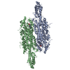


 PDBj
PDBj


 Assembly


 Mass: 154.251 Da / Num. of mol.: 2 / Source method: obtained synthetically / Formula: C4H10O2S2
 Mass: 18.015 Da / Num. of mol.: 1071 / Source method: isolated from a natural source / Formula: H2O
 Sample preparation
 / Beamline: PX9.5 / Wavelength: 0.9798,0.9800,0.9824
 Processing