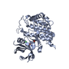
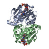
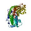
[English] 日本語
Yorodumi
- PDB-3nw5: Crystal structure of insulin-like growth factor 1 receptor (IGF-1... -

+
Open data


ID or keywords:

Loading...

-
Basic information

Entry
Database: PDB / ID: 3nw5
TitleCrystal structure of insulin-like growth factor 1 receptor (IGF-1R-WT) complex with a carbon-linked proline isostere inhibitor (11B)
ComponentsInsulin-like growth factor 1 receptor
KeywordsTRANSFERASE / HORMONE/GROWTH FACTOR / IGF-1R / KINASE DOMAIN
Function / homology
Function and homology information


cardiac atrium development / negative regulation of cholangiocyte apoptotic process / insulin-like growth factor receptor activity / positive regulation of steroid hormone biosynthetic process / protein kinase complex / protein transporter activity / Signaling by Type 1 Insulin-like Growth Factor 1 Receptor (IGF1R) / IRS-related events triggered by IGF1R / insulin-like growth factor binding / negative regulation of muscle cell apoptotic process ...cardiac atrium development / negative regulation of cholangiocyte apoptotic process / insulin-like growth factor receptor activity / positive regulation of steroid hormone biosynthetic process / protein kinase complex / protein transporter activity / Signaling by Type 1 Insulin-like Growth Factor 1 Receptor (IGF1R) / IRS-related events triggered by IGF1R / insulin-like growth factor binding / negative regulation of muscle cell apoptotic process / cellular response to progesterone stimulus / positive regulation of DNA metabolic process / cellular response to zinc ion starvation / cellular response to aldosterone / insulin receptor complex / cellular response to testosterone stimulus / negative regulation of hepatocyte apoptotic process / insulin-like growth factor I binding / insulin receptor activity / transcytosis / alphav-beta3 integrin-IGF-1-IGF1R complex / response to alkaloid / positive regulation of protein-containing complex disassembly / cellular response to angiotensin / cellular response to insulin-like growth factor stimulus / dendritic spine maintenance / response to L-glutamate / insulin binding / establishment of cell polarity / negative regulation of MAPK cascade / positive regulation of axon regeneration / amyloid-beta clearance / positive regulation of cytokinesis / positive regulation of osteoblast proliferation / regulation of JNK cascade / estrous cycle / insulin receptor substrate binding / G-protein alpha-subunit binding / response to vitamin E / SHC-related events triggered by IGF1R / phosphatidylinositol 3-kinase binding / peptidyl-tyrosine autophosphorylation / cellular response to transforming growth factor beta stimulus / T-tubule / cellular response to dexamethasone stimulus / cerebellum development / axonogenesis / insulin-like growth factor receptor signaling pathway / response to nicotine / phosphatidylinositol 3-kinase/protein kinase B signal transduction / cellular response to estradiol stimulus / caveola / cellular response to glucose stimulus / hippocampus development / positive regulation of smooth muscle cell proliferation / insulin receptor binding / receptor protein-tyrosine kinase / cellular response to mechanical stimulus / cellular response to amyloid-beta / cellular senescence / insulin receptor signaling pathway / positive regulation of cold-induced thermogenesis / protein tyrosine kinase activity / response to ethanol / Extra-nuclear estrogen signaling / positive regulation of MAPK cascade / protein autophosphorylation / positive regulation of phosphatidylinositol 3-kinase/protein kinase B signal transduction / receptor complex / positive regulation of cell migration / immune response / axon / intracellular membrane-bounded organelle / neuronal cell body / positive regulation of cell population proliferation / protein-containing complex binding / negative regulation of apoptotic process / signal transduction / ATP binding / membrane / identical protein binding / plasma membrane
Similarity search - Function
Tyrosine-protein kinase, insulin-like receptor / Tyrosine-protein kinase, receptor class II, conserved site / Receptor tyrosine kinase class II signature. / Receptor L-domain / Furin-like cysteine-rich domain / Receptor L-domain superfamily / Furin-like cysteine rich region / Receptor L domain / Furin-like repeat / Furin-like repeats ...Tyrosine-protein kinase, insulin-like receptor / Tyrosine-protein kinase, receptor class II, conserved site / Receptor tyrosine kinase class II signature. / Receptor L-domain / Furin-like cysteine-rich domain / Receptor L-domain superfamily / Furin-like cysteine rich region / Receptor L domain / Furin-like repeat / Furin-like repeats / Growth factor receptor cysteine-rich domain superfamily / Fibronectin type III domain / Fibronectin type 3 domain / Fibronectin type-III domain profile. / Fibronectin type III / Fibronectin type III superfamily / Tyrosine-protein kinase, catalytic domain / Tyrosine kinase, catalytic domain / Tyrosine protein kinases specific active-site signature. / Tyrosine-protein kinase, active site / Protein tyrosine and serine/threonine kinase / Serine-threonine/tyrosine-protein kinase, catalytic domain / Transferase(Phosphotransferase) domain 1 / Transferase(Phosphotransferase); domain 1 / Phosphorylase Kinase; domain 1 / Phosphorylase Kinase; domain 1 / Protein kinase, ATP binding site / Protein kinases ATP-binding region signature. / Immunoglobulin-like fold / Protein kinase domain profile. / Protein kinase domain / Protein kinase-like domain superfamily / 2-Layer Sandwich / Orthogonal Bundle / Mainly Alpha / Alpha Beta
Similarity search - Domain/homology
Chem-LGX / Insulin-like growth factor 1 receptor
Similarity search - Component
Biological speciesHomo sapiens (human)
MethodX-RAY DIFFRACTION / MOLECULAR REPLACEMENT / Resolution: 2.14 Å
AuthorsSack, J.S.
CitationJournal: Bioorg.Med.Chem.Lett. / Year: 2010
Title: Proline isosteres in a series of 2,4-disubstituted pyrrolo[1,2-f][1,2,4]triazine inhibitors of IGF-1R kinase and IR kinase.
Authors: Sampognaro, A.J. / Wittman, M.D. / Carboni, J.M. / Chang, C. / Greer, A.F. / Hurlburt, W.W. / Sack, J.S. / Vyas, D.M.
History
DepositionJul 9, 2010Deposition site: RCSB / Processing site: RCSB
Revision 1.0Jul 28, 2010Provider: repository / Type: Initial release
Revision 1.1Jul 13, 2011Group: Version format compliance
Revision 1.2Feb 21, 2024Group: Data collection / Database references / Derived calculations
Category: chem_comp_atom / chem_comp_bond ...chem_comp_atom / chem_comp_bond / database_2 / struct_ref_seq_dif / struct_site
Item: _database_2.pdbx_DOI / _database_2.pdbx_database_accession ..._database_2.pdbx_DOI / _database_2.pdbx_database_accession / _struct_ref_seq_dif.details / _struct_site.pdbx_auth_asym_id / _struct_site.pdbx_auth_comp_id / _struct_site.pdbx_auth_seq_id

-
Structure visualization

Structure viewerMolecule:
MolmilJmol/JSmol

Downloads & links

-
Assembly

Deposited unit
A: Insulin-like growth factor 1 receptor
hetero molecules


Theoretical massNumber of molelcules
Total (without water)35,4742
Polymers35,0411
Non-polymers4321
Water3,693205
1


  • Idetical with deposited unit
  • defined by author&software
TypeNameSymmetry operationNumber
identity operation1_555x,y,z1
MethodPISA
Unit cell
Length a, b, c (Å)106.642, 41.289, 78.183
Angle α, β, γ (deg.)90.00, 120.21, 90.00
Int Tables number5
Space group name H-MC121

-
Components

#1: Protein Insulin-like growth factor 1 receptor / / Insulin-like growth factor I receptor / IGF-I receptor


Mass: 35041.230 Da / Num. of mol.: 1 / Fragment: KINASE DOMAIN (RESIDUES 982-1286)
Source method: isolated from a genetically manipulated source
Source: (gene. exp.) Homo sapiens (human) / Gene: IGF1R / Production host: unidentified baculovirus
References: UniProt: P08069, receptor protein-tyrosine kinase
#2: Chemical ChemComp-LGX / N-(5-cyclopropyl-1H-pyrazol-3-yl)-2-{(2R)-1-[(6-fluoropyridin-3-yl)carbonyl]pyrrolidin-2-yl}pyrrolo[2,1-f][1,2,4]triazin-4-amine


Mass: 432.454 Da / Num. of mol.: 1 / Source method: obtained synthetically / Formula: C22H21FN8O
#3: Water ChemComp-HOH / water / Water


Mass: 18.015 Da / Num. of mol.: 205 / Source method: isolated from a natural source / Formula: H2O

-
Experimental details

-
Experiment

ExperimentMethod: X-RAY DIFFRACTION / Number of used crystals: 1

-
Sample preparation

CrystalDensity Matthews: 2.12 Å3/Da / Density % sol: 42.05 %

-
Data collection

DiffractionMean temperature: 100 K
Diffraction sourceSource: ROTATING ANODE / Type: RIGAKU FR-E DW / Wavelength: 1.54
DetectorType: RIGAKU SATURN 92 / Detector: CCD / Date: Jun 15, 2006 / Details: MICROMAX CONFOCAL
RadiationProtocol: SINGLE WAVELENGTH / Monochromatic (M) / Laue (L): M / Scattering type: x-ray
Radiation wavelengthWavelength: 1.54 Å / Relative weight: 1
ReflectionResolution: 2.14→50 Å / Num. obs: 16126 / % possible obs: 98.4 % / Observed criterion σ(I): 0 / Redundancy: 3.2 % / Biso Wilson estimate: 35.94 Å2 / Rmerge(I) obs: 0.102 / Net I/σ(I): 10.9
Reflection shellResolution: 2.14→2.23 Å / Redundancy: 2.7 % / Rmerge(I) obs: 0.475 / Mean I/σ(I) obs: 2.1 / % possible all: 94.9

-
Processing

Software
NameVersionClassification
BUSTER2.9.3refinement
HKL-2000(DENZO)data reduction
HKL-2000(SCALEPACK)data scaling
RefinementMethod to determine structure: MOLECULAR REPLACEMENT / Resolution: 2.14→26.91 Å / Cor.coef. Fo:Fc: 0.9444 / Cor.coef. Fo:Fc free: 0.9204 / SU R Cruickshank DPI: 0.287 / Cross valid method: THROUGHOUT / σ(F): 0
RfactorNum. reflection% reflectionSelection details
Rfree0.2537 812 5.04 %RANDOM
Rwork0.1933 ---
obs0.1964 16110 97.54 %-
Displacement parametersBiso mean: 35.29 Å2
Baniso -1Baniso -2Baniso -3
1--0.9039 Å20 Å21.1341 Å2
2---4.128 Å20 Å2
3---5.0319 Å2
Refine analyzeLuzzati coordinate error obs: 0.258 Å
Refinement stepCycle: LAST / Resolution: 2.14→26.91 Å
ProteinNucleic acidLigandSolventTotal
Num. atoms2408 0 32 205 2645
Refine LS restraints
Refine-IDTypeDev idealNumberWeight
X-RAY DIFFRACTIONt_bond_d0.0124972
X-RAY DIFFRACTIONt_angle_deg1.1333722
X-RAY DIFFRACTIONt_dihedral_angle_d8802
X-RAY DIFFRACTIONt_incorr_chiral_ct
X-RAY DIFFRACTIONt_pseud_angle
X-RAY DIFFRACTIONt_trig_c_planes652
X-RAY DIFFRACTIONt_gen_planes3575
X-RAY DIFFRACTIONt_it249720
X-RAY DIFFRACTIONt_nbd05
X-RAY DIFFRACTIONt_omega_torsion3.38
X-RAY DIFFRACTIONt_other_torsion19.4
X-RAY DIFFRACTIONt_improper_torsion
X-RAY DIFFRACTIONt_chiral_improper_torsion3025
X-RAY DIFFRACTIONt_sum_occupancies
X-RAY DIFFRACTIONt_utility_distance
X-RAY DIFFRACTIONt_utility_angle
X-RAY DIFFRACTIONt_utility_torsion
X-RAY DIFFRACTIONt_ideal_dist_contact31934
LS refinement shellResolution: 2.14→2.29 Å / Total num. of bins used: 8
RfactorNum. reflection% reflection
Rfree0.2826 141 5.18 %
Rwork0.2076 2581 -
all0.2115 2722 -
obs--97.54 %

+
About Yorodumi

-
News

-
Feb 9, 2022. New format data for meta-information of EMDB entries

New format data for meta-information of EMDB entries

  • Version 3 of the EMDB header file is now the official format.
  • The previous official version 1.9 will be removed from the archive.

Related info.:EMDB header

External links:wwPDB to switch to version 3 of the EMDB data model

-
Aug 12, 2020. Covid-19 info

Covid-19 info

URL: https://pdbj.org/emnavi/covid19.php

New page: Covid-19 featured information page in EM Navigator.

Related info.:Covid-19 info / Mar 5, 2020. Novel coronavirus structure data

+
Mar 5, 2020. Novel coronavirus structure data

Novel coronavirus structure data

Related info.:Yorodumi Speices / Aug 12, 2020. Covid-19 info

External links:COVID-19 featured content - PDBj / Molecule of the Month (242):Coronavirus Proteases

+
Jan 31, 2019. EMDB accession codes are about to change! (news from PDBe EMDB page)

EMDB accession codes are about to change! (news from PDBe EMDB page)

  • The allocation of 4 digits for EMDB accession codes will soon come to an end. Whilst these codes will remain in use, new EMDB accession codes will include an additional digit and will expand incrementally as the available range of codes is exhausted. The current 4-digit format prefixed with “EMD-” (i.e. EMD-XXXX) will advance to a 5-digit format (i.e. EMD-XXXXX), and so on. It is currently estimated that the 4-digit codes will be depleted around Spring 2019, at which point the 5-digit format will come into force.
  • The EM Navigator/Yorodumi systems omit the EMD- prefix.

Related info.:Q: What is EMD? / ID/Accession-code notation in Yorodumi/EM Navigator

External links:EMDB Accession Codes are Changing Soon! / Contact to PDBj

+
Jul 12, 2017. Major update of PDB

Major update of PDB

  • wwPDB released updated PDB data conforming to the new PDBx/mmCIF dictionary.
  • This is a major update changing the version number from 4 to 5, and with Remediation, in which all the entries are updated.
  • In this update, many items about electron microscopy experimental information are reorganized (e.g. em_software).
  • Now, EM Navigator and Yorodumi are based on the updated data.

External links:wwPDB Remediation / Enriched Model Files Conforming to OneDep Data Standards Now Available in the PDB FTP Archive

-
Yorodumi

Thousand views of thousand structures

  • Yorodumi is a browser for structure data from EMDB, PDB, SASBDB, etc.
  • This page is also the successor to EM Navigator detail page, and also detail information page/front-end page for Omokage search.
  • The word "yorodu" (or yorozu) is an old Japanese word meaning "ten thousand". "mi" (miru) is to see.

Related info.:EMDB / PDB / SASBDB / Comparison of 3 databanks / Yorodumi Search / Aug 31, 2016. New EM Navigator & Yorodumi / Yorodumi Papers / Jmol/JSmol / Function and homology information / Changes in new EM Navigator and Yorodumi

Read more