 Movie
Movie Controller

[English] 日本語
 Yorodumi
- PDB-6add: The crystal structure of Rv2747 from Mycobacterium tuberculosis i... -
+ Open data

- Basic information
| Entry | Database: PDB / ID: 6add | ||||||
|---|---|---|---|---|---|---|---|
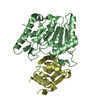
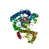
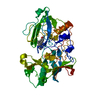
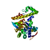
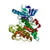
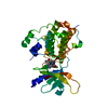
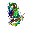
| Title | The crystal structure of Rv2747 from Mycobacterium tuberculosis in complex with CoA and NLQ | ||||||
 Components Components | Amino-acid acetyltransferase | ||||||
 Keywords Keywords |  TRANSFERASE / Acetyl transferase / Mycobacterium tuberculosis / Rv2747 TRANSFERASE / Acetyl transferase / Mycobacterium tuberculosis / Rv2747 | ||||||
| Function / homology |  Function and homology information Function and homology informationmethione N-acyltransferase activity / amino-acid N-acetyltransferase / acetyl-CoA:L-glutamate N-acetyltransferase activity / arginine biosynthetic process via ornithine / N-acetyltransferase activity / protein homotetramerization / protein homodimerization activity / cytoplasmSimilarity search - Function | ||||||
| Biological species |  Mycobacterium tuberculosis H37Rv (bacteria) Mycobacterium tuberculosis H37Rv (bacteria) | ||||||
| Method | X-RAY DIFFRACTION / MOLECULAR REPLACEMENT / molecular replacement / Resolution: 2.301 Å | ||||||
 Authors Authors | Das, U. / Singh, E. / Pal, R.K. / Tiruttani Subhramanyam, U.K. / Gourinath, S. / Srinivasan, A. | ||||||
| Funding support |  India, 1items India, 1items
| ||||||
 Citation Citation | Journal: Int. J. Biol. Macromol. / Year: 2019 Title: Structural insights into the substrate binding mechanism of novel ArgA from Mycobacterium tuberculosis Authors: Das, U. / Singh, E. / Dharavath, S. / Tiruttani Subhramanyam, U.K. / Pal, R.K. / Vijayan, R. / Menon, S. / Kumar, S. / Gourinath, S. / Srinivasan, A. | ||||||
| History |
|
- Structure visualization
| Structure viewer | Molecule: MolmilJmol/JSmol |
|---|
- Downloads & links
-Download
| PDBx/mmCIF format | 6add.cif.gz | 157.9 KB | Display | PDBx/mmCIF format |
|---|---|---|---|---|
| PDB format | pdb6add.ent.gz | 123.2 KB | Display | PDB format |
| PDBx/mmJSON format | 6add.json.gz | Tree view | PDBx/mmJSON format | |
| Others |  Other downloads Other downloads |
-Validation report
| Arichive directory | https://data.pdbj.org/pub/pdb/validation_reports/ad/6addftp://data.pdbj.org/pub/pdb/validation_reports/ad/6add | HTTPS FTP |
|---|
-Related structure data
| Related structure data |  5yo2SC S: Starting model for refinement C: citing same article ( |
|---|---|
| Similar structure data |


-Links
 PDBj
PDBj


- Assembly
| Deposited unit | 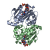
| ||||||||||||||||||||||||||||||||||||||||||||||||||||||||||||||||||||||||||||||||||||||||||
|---|---|---|---|---|---|---|---|---|---|---|---|---|---|---|---|---|---|---|---|---|---|---|---|---|---|---|---|---|---|---|---|---|---|---|---|---|---|---|---|---|---|---|---|---|---|---|---|---|---|---|---|---|---|---|---|---|---|---|---|---|---|---|---|---|---|---|---|---|---|---|---|---|---|---|---|---|---|---|---|---|---|---|---|---|---|---|---|---|---|---|---|
| 1 |
| ||||||||||||||||||||||||||||||||||||||||||||||||||||||||||||||||||||||||||||||||||||||||||
| Unit cell |
| ||||||||||||||||||||||||||||||||||||||||||||||||||||||||||||||||||||||||||||||||||||||||||
| Noncrystallographic symmetry (NCS) | NCS domain:
NCS domain segments: Ens-ID: 1
|