 Movie
Movie Controller

[English] 日本語
 Yorodumi
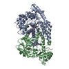
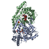
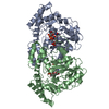
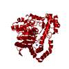
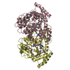
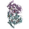
- PDB-3nu7: WbpE, an Aminotransferase from Pseudomonas aeruginosa Involved in... -
+ Open data

- Basic information
| Entry | Database: PDB / ID: 3nu7 | ||||||
|---|---|---|---|---|---|---|---|
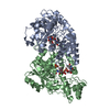
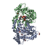
| Title | WbpE, an Aminotransferase from Pseudomonas aeruginosa Involved in O-antigen Assembly in Complex with the Cofactor PMP | ||||||
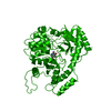
 Components Components | Aminotransferase WbpE | ||||||
 Keywords Keywords |  TRANSFERASE / Domain-swapped beta hairpin / Aminotransferase / PLP / PMP / UDP-GlcNAc(3NH2)A / Lipopolysaccharide / O-antigen / B-band TRANSFERASE / Domain-swapped beta hairpin / Aminotransferase / PLP / PMP / UDP-GlcNAc(3NH2)A / Lipopolysaccharide / O-antigen / B-band | ||||||
| Function / homology |  Function and homology informationUDP-2-acetamido-2-deoxy-ribo-hexuluronate aminotransferase / UDP-glucuronate biosynthetic process / O antigen biosynthetic process / polysaccharide biosynthetic process / lipopolysaccharide biosynthetic process / transaminase activity / cell wall organization / pyridoxal phosphate binding Function and homology informationUDP-2-acetamido-2-deoxy-ribo-hexuluronate aminotransferase / UDP-glucuronate biosynthetic process / O antigen biosynthetic process / polysaccharide biosynthetic process / lipopolysaccharide biosynthetic process / transaminase activity / cell wall organization / pyridoxal phosphate bindingSimilarity search - Function | ||||||
| Biological species |   Pseudomonas aeruginosa (bacteria) Pseudomonas aeruginosa (bacteria) | ||||||
| Method | X-RAY DIFFRACTION / SYNCHROTRON / MOLECULAR REPLACEMENT / Resolution: 1.95 Å | ||||||
 Authors Authors | Larkin, A. / Olivier, N.B. / Imperiali, B. | ||||||
 Citation Citation | Journal: Biochemistry / Year: 2010 Title: Structural Analysis of WbpE from Pseudomonas aeruginosa PAO1: A Nucleotide Sugar Aminotransferase Involved in O-Antigen Assembly Authors: Larkin, A. / Olivier, N.B. / Imperiali, B. | ||||||
| History |
|
- Structure visualization
| Structure viewer | Molecule: MolmilJmol/JSmol |
|---|
- Downloads & links
-Download
| PDBx/mmCIF format | 3nu7.cif.gz | 152.3 KB | Display | PDBx/mmCIF format |
|---|---|---|---|---|
| PDB format | pdb3nu7.ent.gz | 120.6 KB | Display | PDB format |
| PDBx/mmJSON format | 3nu7.json.gz | Tree view | PDBx/mmJSON format | |
| Others |  Other downloads Other downloads |
-Validation report
| Arichive directory | https://data.pdbj.org/pub/pdb/validation_reports/nu/3nu7ftp://data.pdbj.org/pub/pdb/validation_reports/nu/3nu7 | HTTPS FTP |
|---|
-Related structure data


-Links
 PDBj
PDBj- Assembly
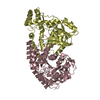
| Deposited unit | 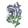
| ||||||||
|---|---|---|---|---|---|---|---|---|---|
| 1 |
| ||||||||
| Unit cell |
|
-Components
| #1: Protein | Mass: 38965.547 Da / Num. of mol.: 2 Source method: isolated from a genetically manipulated source Source: (gene. exp.) Pseudomonas aeruginosa (bacteria) / Strain: PAO1-LAC / Gene: PA3155, wbpE / Plasmid: pET32a / Production host: Escherichia coli (E. coli) / Strain (production host): BL21(DE3) RIL / References: UniProt: Q9HZ76#2: Chemical | Glycerol  Mass: 92.094 Da / Num. of mol.: 3 / Source method: obtained synthetically / Formula: C3H8O3 Mass: 92.094 Da / Num. of mol.: 3 / Source method: obtained synthetically / Formula: C3H8O3#3: Chemical |   Mass: 248.173 Da / Num. of mol.: 2 / Source method: obtained synthetically / Formula: C8H13N2O5P Mass: 248.173 Da / Num. of mol.: 2 / Source method: obtained synthetically / Formula: C8H13N2O5P#4: Water | ChemComp-HOH / | Water Mass: 18.015 Da / Num. of mol.: 377 / Source method: isolated from a natural source / Formula: H2O Mass: 18.015 Da / Num. of mol.: 377 / Source method: isolated from a natural source / Formula: H2O |
|---|
-Experimental details
-Experiment
| Experiment | Method: X-RAY DIFFRACTION / Number of used crystals: 1 |
|---|
- Sample preparation
| Crystal | Density Matthews: 2.06 Å3/Da / Density % sol: 40.23 % |
|---|---|
| Crystal grow | Temperature: 298 K / Method: vapor diffusion, hanging drop / pH: 5.5 Details: Crystallization conditions: 0.1 M Bis-Tris, pH 5.5, 0.2 M Ammonium sulfate, 25% PEG 3350 in the reservoir; Protein solution contains 25 mM HEPES, pH 8.0, 100 mM NaCl, 0.5% glycerol; Drop ...Details: Crystallization conditions: 0.1 M Bis-Tris, pH 5.5, 0.2 M Ammonium sulfate, 25% PEG 3350 in the reservoir; Protein solution contains 25 mM HEPES, pH 8.0, 100 mM NaCl, 0.5% glycerol; Drop made by mixing 1.5 uL of protein and reservoir solutions., VAPOR DIFFUSION, HANGING DROP, temperature 298K |
-Data collection
| Diffraction | Mean temperature: 110 K |
|---|---|
| Diffraction source | Source: SYNCHROTRON / Site: NSLS  / Beamline: X6A / Beamline: X6A |
| Detector | Type: ADSC QUANTUM 270 / Detector: CCD / Date: Jun 26, 2009 / Details: Toroidal Focusing Mirror |
| Radiation | Monochromator: SI(111) Channel Cut Monochromator / Protocol: SINGLE WAVELENGTH / Monochromatic (M) / Laue (L): M / Scattering type: x-ray |
| Radiation wavelength | Relative weight: 1 |
| Reflection | Resolution: 1.95→50 Å / Num. obs: 45308 / % possible obs: 99.8 % / Redundancy: 14.5 % / Rmerge(I) obs: 0.073 / Net I/σ(I): 53.4 |
| Reflection shell | Resolution: 1.95→1.98 Å / Redundancy: 14.5 % / % possible all: 100 |
- Processing
| Software |
| |||||||||||||||||||||||||||||||||||||||||||||||||||||||||||||||||
|---|---|---|---|---|---|---|---|---|---|---|---|---|---|---|---|---|---|---|---|---|---|---|---|---|---|---|---|---|---|---|---|---|---|---|---|---|---|---|---|---|---|---|---|---|---|---|---|---|---|---|---|---|---|---|---|---|---|---|---|---|---|---|---|---|---|---|
| Refinement | Method to determine structure: MOLECULAR REPLACEMENT Starting model: SeMet Derivative Resolution: 1.95→34.66 Å / Cor.coef. Fo:Fc: 0.951 / Cor.coef. Fo:Fc free: 0.93 / SU B: 4.117 / SU ML: 0.12 / Cross valid method: THROUGHOUT / ESU R Free: 0.175 / Stereochemistry target values: MAXIMUM LIKELIHOOD / Details: HYDROGENS HAVE BEEN ADDED IN THE RIDING POSITIONS
| |||||||||||||||||||||||||||||||||||||||||||||||||||||||||||||||||
| Solvent computation | Ion probe radii: 0.8 Å / Shrinkage radii: 0.8 Å / VDW probe radii: 1.4 Å / Solvent model: MASK | |||||||||||||||||||||||||||||||||||||||||||||||||||||||||||||||||
| Displacement parameters | Biso mean: 28.367 Å2
| |||||||||||||||||||||||||||||||||||||||||||||||||||||||||||||||||
| Refinement step | Cycle: LAST / Resolution: 1.95→34.66 Å
| |||||||||||||||||||||||||||||||||||||||||||||||||||||||||||||||||
| Refine LS restraints |
| |||||||||||||||||||||||||||||||||||||||||||||||||||||||||||||||||
| LS refinement shell | Resolution: 1.949→1.999 Å / Total num. of bins used: 20
|
