 Movie
Movie Controller

[English] 日本語
 Yorodumi
- PDB-3ns8: Crystal structure of an open conformation of Lys48-linked diubiqu... -
+ Open data

- Basic information
| Entry | Database: PDB / ID: 3ns8 | ||||||
|---|---|---|---|---|---|---|---|
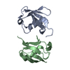
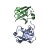
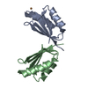
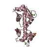
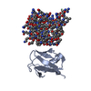
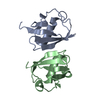
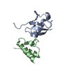
| Title | Crystal structure of an open conformation of Lys48-linked diubiquitin at pH 7.5 | ||||||
 Components Components | Ubiquitin | ||||||
 Keywords Keywords | SIGNALING PROTEIN / Diubiquitin / isopeptide bond | ||||||
| Function / homology |  Function and homology information: / : / protein modification process => GO:0036211 / Peptide chain elongation / Selenocysteine synthesis / Formation of a pool of free 40S subunits / Eukaryotic Translation Termination / Response of EIF2AK4 (GCN2) to amino acid deficiency / SRP-dependent cotranslational protein targeting to membrane / Viral mRNA Translation ...: / : / protein modification process => GO:0036211 / Peptide chain elongation / Selenocysteine synthesis / Formation of a pool of free 40S subunits / Eukaryotic Translation Termination / Response of EIF2AK4 (GCN2) to amino acid deficiency / SRP-dependent cotranslational protein targeting to membrane / Viral mRNA Translation / Nonsense Mediated Decay (NMD) independent of the Exon Junction Complex (EJC) / GTP hydrolysis and joining of the 60S ribosomal subunit / L13a-mediated translational silencing of Ceruloplasmin expression / Major pathway of rRNA processing in the nucleolus and cytosol / Nonsense Mediated Decay (NMD) enhanced by the Exon Junction Complex (EJC) / Maturation of protein E / Maturation of protein E / ER Quality Control Compartment (ERQC) / Myoclonic epilepsy of Lafora / FLT3 signaling by CBL mutants / Prevention of phagosomal-lysosomal fusion / IRAK2 mediated activation of TAK1 complex / Alpha-protein kinase 1 signaling pathway / Glycogen synthesis / IRAK1 recruits IKK complex / IRAK1 recruits IKK complex upon TLR7/8 or 9 stimulation / Membrane binding and targetting of GAG proteins / Constitutive Signaling by NOTCH1 HD Domain Mutants / Endosomal Sorting Complex Required For Transport (ESCRT) / NOTCH2 Activation and Transmission of Signal to the Nucleus / IRAK2 mediated activation of TAK1 complex upon TLR7/8 or 9 stimulation / PTK6 Regulates RTKs and Their Effectors AKT1 and DOK1 / Negative regulation of FLT3 / Regulation of FZD by ubiquitination / TICAM1,TRAF6-dependent induction of TAK1 complex / TICAM1-dependent activation of IRF3/IRF7 / APC/C:Cdc20 mediated degradation of Cyclin B / p75NTR recruits signalling complexes / Downregulation of ERBB4 signaling / TRAF6 mediated IRF7 activation in TLR7/8 or 9 signaling / APC-Cdc20 mediated degradation of Nek2A / PINK1-PRKN Mediated Mitophagy / TRAF6-mediated induction of TAK1 complex within TLR4 complex / Pexophagy / InlA-mediated entry of Listeria monocytogenes into host cells / Regulation of innate immune responses to cytosolic DNA / VLDLR internalisation and degradation / Downregulation of ERBB2:ERBB3 signaling / NRIF signals cell death from the nucleus / Activated NOTCH1 Transmits Signal to the Nucleus / NF-kB is activated and signals survival / Regulation of PTEN localization / Regulation of BACH1 activity / Translesion synthesis by REV1 / Synthesis of active ubiquitin: roles of E1 and E2 enzymes / Translesion synthesis by POLK / MAP3K8 (TPL2)-dependent MAPK1/3 activation / cytosolic ribosome / TICAM1, RIP1-mediated IKK complex recruitment / Gap-filling DNA repair synthesis and ligation in GG-NER / Downregulation of TGF-beta receptor signaling / Translesion synthesis by POLI / Josephin domain DUBs / Activation of IRF3, IRF7 mediated by TBK1, IKKε (IKBKE) / Regulation of activated PAK-2p34 by proteasome mediated degradation / InlB-mediated entry of Listeria monocytogenes into host cell / IKK complex recruitment mediated by RIP1 / JNK (c-Jun kinases) phosphorylation and activation mediated by activated human TAK1 / TGF-beta receptor signaling in EMT (epithelial to mesenchymal transition) / N-glycan trimming in the ER and Calnexin/Calreticulin cycle / Autodegradation of Cdh1 by Cdh1:APC/C / TNFR1-induced NF-kappa-B signaling pathway / APC/C:Cdc20 mediated degradation of Securin / Asymmetric localization of PCP proteins / TCF dependent signaling in response to WNT / SCF-beta-TrCP mediated degradation of Emi1 / NIK-->noncanonical NF-kB signaling / Regulation of NF-kappa B signaling / Ubiquitin-dependent degradation of Cyclin D / AUF1 (hnRNP D0) binds and destabilizes mRNA / NOTCH3 Activation and Transmission of Signal to the Nucleus / TNFR2 non-canonical NF-kB pathway / activated TAK1 mediates p38 MAPK activation / Assembly of the pre-replicative complex / Vpu mediated degradation of CD4 / Negative regulators of DDX58/IFIH1 signaling / Degradation of DVL / Deactivation of the beta-catenin transactivating complex / Ubiquitin Mediated Degradation of Phosphorylated Cdc25A / Regulation of signaling by CBL / Dectin-1 mediated noncanonical NF-kB signaling / Hh mutants are degraded by ERAD / Cdc20:Phospho-APC/C mediated degradation of Cyclin A / Recognition of DNA damage by PCNA-containing replication complex / Fanconi Anemia Pathway / Negative regulation of FGFR3 signaling / Peroxisomal protein import / Degradation of AXIN / Downregulation of SMAD2/3:SMAD4 transcriptional activity / Defective CFTR causes cystic fibrosis Function and homology information: / : / protein modification process => GO:0036211 / Peptide chain elongation / Selenocysteine synthesis / Formation of a pool of free 40S subunits / Eukaryotic Translation Termination / Response of EIF2AK4 (GCN2) to amino acid deficiency / SRP-dependent cotranslational protein targeting to membrane / Viral mRNA Translation ...: / : / protein modification process => GO:0036211 / Peptide chain elongation / Selenocysteine synthesis / Formation of a pool of free 40S subunits / Eukaryotic Translation Termination / Response of EIF2AK4 (GCN2) to amino acid deficiency / SRP-dependent cotranslational protein targeting to membrane / Viral mRNA Translation / Nonsense Mediated Decay (NMD) independent of the Exon Junction Complex (EJC) / GTP hydrolysis and joining of the 60S ribosomal subunit / L13a-mediated translational silencing of Ceruloplasmin expression / Major pathway of rRNA processing in the nucleolus and cytosol / Nonsense Mediated Decay (NMD) enhanced by the Exon Junction Complex (EJC) / Maturation of protein E / Maturation of protein E / ER Quality Control Compartment (ERQC) / Myoclonic epilepsy of Lafora / FLT3 signaling by CBL mutants / Prevention of phagosomal-lysosomal fusion / IRAK2 mediated activation of TAK1 complex / Alpha-protein kinase 1 signaling pathway / Glycogen synthesis / IRAK1 recruits IKK complex / IRAK1 recruits IKK complex upon TLR7/8 or 9 stimulation / Membrane binding and targetting of GAG proteins / Constitutive Signaling by NOTCH1 HD Domain Mutants / Endosomal Sorting Complex Required For Transport (ESCRT) / NOTCH2 Activation and Transmission of Signal to the Nucleus / IRAK2 mediated activation of TAK1 complex upon TLR7/8 or 9 stimulation / PTK6 Regulates RTKs and Their Effectors AKT1 and DOK1 / Negative regulation of FLT3 / Regulation of FZD by ubiquitination / TICAM1,TRAF6-dependent induction of TAK1 complex / TICAM1-dependent activation of IRF3/IRF7 / APC/C:Cdc20 mediated degradation of Cyclin B / p75NTR recruits signalling complexes / Downregulation of ERBB4 signaling / TRAF6 mediated IRF7 activation in TLR7/8 or 9 signaling / APC-Cdc20 mediated degradation of Nek2A / PINK1-PRKN Mediated Mitophagy / TRAF6-mediated induction of TAK1 complex within TLR4 complex / Pexophagy / InlA-mediated entry of Listeria monocytogenes into host cells / Regulation of innate immune responses to cytosolic DNA / VLDLR internalisation and degradation / Downregulation of ERBB2:ERBB3 signaling / NRIF signals cell death from the nucleus / Activated NOTCH1 Transmits Signal to the Nucleus / NF-kB is activated and signals survival / Regulation of PTEN localization / Regulation of BACH1 activity / Translesion synthesis by REV1 / Synthesis of active ubiquitin: roles of E1 and E2 enzymes / Translesion synthesis by POLK / MAP3K8 (TPL2)-dependent MAPK1/3 activation / cytosolic ribosome / TICAM1, RIP1-mediated IKK complex recruitment / Gap-filling DNA repair synthesis and ligation in GG-NER / Downregulation of TGF-beta receptor signaling / Translesion synthesis by POLI / Josephin domain DUBs / Activation of IRF3, IRF7 mediated by TBK1, IKKε (IKBKE) / Regulation of activated PAK-2p34 by proteasome mediated degradation / InlB-mediated entry of Listeria monocytogenes into host cell / IKK complex recruitment mediated by RIP1 / JNK (c-Jun kinases) phosphorylation and activation mediated by activated human TAK1 / TGF-beta receptor signaling in EMT (epithelial to mesenchymal transition) / N-glycan trimming in the ER and Calnexin/Calreticulin cycle / Autodegradation of Cdh1 by Cdh1:APC/C / TNFR1-induced NF-kappa-B signaling pathway / APC/C:Cdc20 mediated degradation of Securin / Asymmetric localization of PCP proteins / TCF dependent signaling in response to WNT / SCF-beta-TrCP mediated degradation of Emi1 / NIK-->noncanonical NF-kB signaling / Regulation of NF-kappa B signaling / Ubiquitin-dependent degradation of Cyclin D / AUF1 (hnRNP D0) binds and destabilizes mRNA / NOTCH3 Activation and Transmission of Signal to the Nucleus / TNFR2 non-canonical NF-kB pathway / activated TAK1 mediates p38 MAPK activation / Assembly of the pre-replicative complex / Vpu mediated degradation of CD4 / Negative regulators of DDX58/IFIH1 signaling / Degradation of DVL / Deactivation of the beta-catenin transactivating complex / Ubiquitin Mediated Degradation of Phosphorylated Cdc25A / Regulation of signaling by CBL / Dectin-1 mediated noncanonical NF-kB signaling / Hh mutants are degraded by ERAD / Cdc20:Phospho-APC/C mediated degradation of Cyclin A / Recognition of DNA damage by PCNA-containing replication complex / Fanconi Anemia Pathway / Negative regulation of FGFR3 signaling / Peroxisomal protein import / Degradation of AXIN / Downregulation of SMAD2/3:SMAD4 transcriptional activity / Defective CFTR causes cystic fibrosisSimilarity search - Function | ||||||
| Biological species |  Homo sapiens (human) Homo sapiens (human) | ||||||
| Method | X-RAY DIFFRACTION / SYNCHROTRON / MOLECULAR REPLACEMENT / Resolution: 1.707 Å | ||||||
 Authors Authors | Zhang, D. / LaRonde-LeBlanc, N. / Fushman, D. | ||||||
 Citation Citation | Journal: Biochim.Biophys.Acta / Year: 2012 Title: Structural and biochemical studies of the open state of Lys48-linked diubiquitin. Authors: Lai, M.Y. / Zhang, D. / Laronde-Leblanc, N. / Fushman, D. | ||||||
| History |
|
- Structure visualization
| Structure viewer | Molecule: MolmilJmol/JSmol |
|---|
- Downloads & links
-Download
| PDBx/mmCIF format | 3ns8.cif.gz | 47.2 KB | Display | PDBx/mmCIF format |
|---|---|---|---|---|
| PDB format | pdb3ns8.ent.gz | 32.7 KB | Display | PDB format |
| PDBx/mmJSON format | 3ns8.json.gz | Tree view | PDBx/mmJSON format | |
| Others |  Other downloads Other downloads |
-Validation report
| Arichive directory | https://data.pdbj.org/pub/pdb/validation_reports/ns/3ns8ftp://data.pdbj.org/pub/pdb/validation_reports/ns/3ns8 | HTTPS FTP |
|---|
-Related structure data
| Related structure data |  1ubqS S: Starting model for refinement |
|---|---|
| Similar structure data |


-Links
 PDBj
PDBj


- Assembly
| Deposited unit | 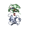
| ||||||||
|---|---|---|---|---|---|---|---|---|---|
| 1 |
| ||||||||
| 2 | 
| ||||||||
| 3 | 
| ||||||||
| Unit cell |
| ||||||||
| Details | The biological unit is two monomers linked by an isopeptide bond between the carboxy-terminal glycine 76 of one monomer, and the lysine 48 side chain of the second monomer. |
-Components
| #1: Protein | Mass: 8576.831 Da / Num. of mol.: 2 Source method: isolated from a genetically manipulated source Source: (gene. exp.) Homo sapiens (human) / Gene: RPS27A / Production host:  Escherichia coli (E. coli) / Strain (production host): BL21 / References: UniProt: P62988, UniProt: P0CG48*PLUS Escherichia coli (E. coli) / Strain (production host): BL21 / References: UniProt: P62988, UniProt: P0CG48*PLUS#2: Chemical | Glycerol  Mass: 92.094 Da / Num. of mol.: 2 / Source method: obtained synthetically / Formula: C3H8O3 Mass: 92.094 Da / Num. of mol.: 2 / Source method: obtained synthetically / Formula: C3H8O3#3: Chemical | ChemComp-EPE / | HEPES  Mass: 238.305 Da / Num. of mol.: 1 / Source method: obtained synthetically / Formula: C8H18N2O4S / Comment: pH buffer*YM Mass: 238.305 Da / Num. of mol.: 1 / Source method: obtained synthetically / Formula: C8H18N2O4S / Comment: pH buffer*YM#4: Water | ChemComp-HOH / | Water Mass: 18.015 Da / Num. of mol.: 120 / Source method: isolated from a natural source / Formula: H2O Mass: 18.015 Da / Num. of mol.: 120 / Source method: isolated from a natural source / Formula: H2O |
|---|
-Experimental details
-Experiment
| Experiment | Method: X-RAY DIFFRACTION / Number of used crystals: 1 |
|---|
- Sample preparation
| Crystal | Density Matthews: 1.85 Å3/Da / Density % sol: 33.58 % |
|---|---|
| Crystal grow | Temperature: 293 K / Method: vapor diffusion, sitting drop / pH: 7.5 Details: HEPES, PEG 8000, Ethylene Glycol, pH 7.5, VAPOR DIFFUSION, SITTING DROP, temperature 293K |
-Data collection
| Diffraction | Mean temperature: 100 K |
|---|---|
| Diffraction source | Source: SYNCHROTRON / Site: APS  / Beamline: 24-ID-E / Wavelength: 0.97927 Å / Beamline: 24-ID-E / Wavelength: 0.97927 Å |
| Detector | Type: ADSC QUANTUM 315 / Detector: CCD / Date: Jun 14, 2007 |
| Radiation | Monochromator: single crystal Si(111) side bounce monochromator Protocol: SINGLE WAVELENGTH / Monochromatic (M) / Laue (L): M / Scattering type: x-ray |
| Radiation wavelength | Wavelength: 0.97927 Å / Relative weight: 1 |
| Reflection | Resolution: 1.707→18.3 Å / Num. all: 13670 / Num. obs: 12906 / % possible obs: 94.41 % / Observed criterion σ(F): 0 / Observed criterion σ(I): 2 / Redundancy: 4.5 % / Biso Wilson estimate: 17.44 Å2 / Rsym value: 0.073 / Net I/σ(I): 17.3 |
| Reflection shell | Resolution: 1.71→1.76 Å / Redundancy: 3.7 % / Mean I/σ(I) obs: 5.4 / Num. unique all: 968 / Rsym value: 0.18 / % possible all: 72.7 |
- Processing
| Software |
| ||||||||||||||||||||||||||||||||||||||||||
|---|---|---|---|---|---|---|---|---|---|---|---|---|---|---|---|---|---|---|---|---|---|---|---|---|---|---|---|---|---|---|---|---|---|---|---|---|---|---|---|---|---|---|---|
| Refinement | Method to determine structure: MOLECULAR REPLACEMENT Starting model: 1UBQ Resolution: 1.707→18.293 Å / SU ML: 0.22 / Isotropic thermal model: Isotropic / Cross valid method: THROUGHOUT / σ(F): 1.36 / Stereochemistry target values: ML
| ||||||||||||||||||||||||||||||||||||||||||
| Solvent computation | Shrinkage radii: 0.9 Å / VDW probe radii: 1.11 Å / Solvent model: FLAT BULK SOLVENT MODEL / Bsol: 65.935 Å2 / ksol: 0.406 e/Å3 | ||||||||||||||||||||||||||||||||||||||||||
| Displacement parameters | Biso mean: 21.1 Å2
| ||||||||||||||||||||||||||||||||||||||||||
| Refine analyze | Luzzati coordinate error obs: 0.22 Å | ||||||||||||||||||||||||||||||||||||||||||
| Refinement step | Cycle: LAST / Resolution: 1.707→18.293 Å
| ||||||||||||||||||||||||||||||||||||||||||
| Refine LS restraints |
| ||||||||||||||||||||||||||||||||||||||||||
| LS refinement shell |
|
