 Movie
Movie Controller


+ Open data

- Basic information
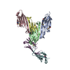
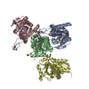
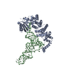
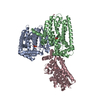
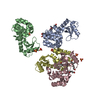
| Entry | Database: PDB / ID: 3now | ||||||
|---|---|---|---|---|---|---|---|
| Title | UNC-45 from Drosophila melanogaster | ||||||
 Components Components | UNC-45 protein, SD10334p | ||||||
 Keywords Keywords |  PROTEIN BINDING / Armadillo Repeat / Hsp90 / Myosin / Tetra-tricopeptide Repeat / TPR / binding protein required for myosin function PROTEIN BINDING / Armadillo Repeat / Hsp90 / Myosin / Tetra-tricopeptide Repeat / TPR / binding protein required for myosin function | ||||||
| Function / homology |  Function and homology information Function and homology informationsomatic muscle development / myosin filament assembly / muscle organ development / chaperone-mediated protein folding / negative regulation of innate immune response / Hsp90 protein binding / cellular response to heat / defense response to Gram-negative bacterium / cell differentiation / cytoplasmSimilarity search - Function | ||||||
| Biological species |  Drosophila melanogaster (fruit fly) Drosophila melanogaster (fruit fly) | ||||||
| Method | X-RAY DIFFRACTION / SYNCHROTRON / SAD / Resolution: 2.992 Å | ||||||
 Authors Authors | Lee, C.F. / Hauenstein, A.V. / Fleming, J.K. / Gasper, W.C. / Engelke, V. / Banumathi, S. / Bernstein, S.I. / Huxford, T. | ||||||
 Citation Citation | Journal: Structure / Year: 2011 Title: X-ray Crystal Structure of the UCS Domain-Containing UNC-45 Myosin Chaperone from Drosophila melanogaster. Authors: Lee, C.F. / Hauenstein, A.V. / Fleming, J.K. / Gasper, W.C. / Engelke, V. / Sankaran, B. / Bernstein, S.I. / Huxford, T. | ||||||
| History |
|
- Structure visualization
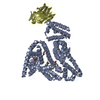
| Structure viewer | Molecule: MolmilJmol/JSmol |
|---|
- Downloads & links
-Download
| PDBx/mmCIF format | 3now.cif.gz | 161.5 KB | Display | PDBx/mmCIF format |
|---|---|---|---|---|
| PDB format | pdb3now.ent.gz | 127 KB | Display | PDB format |
| PDBx/mmJSON format | 3now.json.gz | Tree view | PDBx/mmJSON format | |
| Others |  Other downloads Other downloads |
-Validation report
| Arichive directory | https://data.pdbj.org/pub/pdb/validation_reports/no/3nowftp://data.pdbj.org/pub/pdb/validation_reports/no/3now | HTTPS FTP |
|---|
-Related structure data
| Similar structure data |
|---|


-Links
 PDBj
PDBj


- Assembly
| Deposited unit | 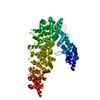
| ||||||||
|---|---|---|---|---|---|---|---|---|---|
| 1 |
| ||||||||
| 2 | 
| ||||||||
| Unit cell |
|
-Components
| #1: Protein | Mass: 89813.523 Da / Num. of mol.: 1 / Fragment: UNP residues 138-947 Source method: isolated from a genetically manipulated source Source: (gene. exp.) Drosophila melanogaster (fruit fly) / Gene: CG2708, Tom34 / Plasmid: pET16b / Production host:  Escherichia coli (E. coli) / Strain (production host): Rosetta (DE3) pLysS / References: UniProt: Q960B1 Escherichia coli (E. coli) / Strain (production host): Rosetta (DE3) pLysS / References: UniProt: Q960B1 |
|---|
-Experimental details
-Experiment
| Experiment | Method: X-RAY DIFFRACTION / Number of used crystals: 1 |
|---|
- Sample preparation
| Crystal | Density Matthews: 5.79 Å3/Da / Density % sol: 78.7 % |
|---|---|
| Crystal grow | Temperature: 291 K / Method: vapor diffusion, hanging drop / pH: 5.6 Details: 0.1 M sodium citrate, 0.2 M ammonium acetate, 25% (w/v) PEG 4000, 1% (v/v) ethylene glycol, pH 5.6, VAPOR DIFFUSION, HANGING DROP, temperature 291.0K |
-Data collection
| Diffraction | Mean temperature: 100 K |
|---|---|
| Diffraction source | Source: SYNCHROTRON / Site: ALS  / Beamline: 8.2.2 / Wavelength: 1 Å / Beamline: 8.2.2 / Wavelength: 1 Å |
| Detector | Type: ADSC QUANTUM 315 / Detector: CCD / Date: Aug 10, 2007 |
| Radiation | Scattering type: x-ray |
| Radiation wavelength | Wavelength: 1 Å / Relative weight: 1 |
| Reflection | Resolution: 2.992→50 Å / Num. obs: 42099 / % possible obs: 99.8 % / Observed criterion σ(F): 2 / Observed criterion σ(I): 2 / Redundancy: 9.5 % / Rsym value: 0.0115 / Net I/σ(I): 19.2 |
| Reflection shell | Resolution: 2.992→3.11 Å / Redundancy: 6.5 % / Num. unique all: 4113 / % possible all: 97.8 |
- Processing
| Software |
| |||||||||||||||||||||||||||||||||||||||||||||||||||||||||||||||||||||||||||||
|---|---|---|---|---|---|---|---|---|---|---|---|---|---|---|---|---|---|---|---|---|---|---|---|---|---|---|---|---|---|---|---|---|---|---|---|---|---|---|---|---|---|---|---|---|---|---|---|---|---|---|---|---|---|---|---|---|---|---|---|---|---|---|---|---|---|---|---|---|---|---|---|---|---|---|---|---|---|---|
| Refinement | Method to determine structure: SAD / Resolution: 2.992→49.197 Å / SU ML: 0.45 / σ(F): 0.15 / Stereochemistry target values: ML
| |||||||||||||||||||||||||||||||||||||||||||||||||||||||||||||||||||||||||||||
| Solvent computation | Shrinkage radii: 0.9 Å / VDW probe radii: 1.11 Å / Solvent model: FLAT BULK SOLVENT MODEL / Bsol: 20.07 Å2 / ksol: 0.292 e/Å3 | |||||||||||||||||||||||||||||||||||||||||||||||||||||||||||||||||||||||||||||
| Displacement parameters |
| |||||||||||||||||||||||||||||||||||||||||||||||||||||||||||||||||||||||||||||
| Refinement step | Cycle: LAST / Resolution: 2.992→49.197 Å
| |||||||||||||||||||||||||||||||||||||||||||||||||||||||||||||||||||||||||||||
| Refine LS restraints |
| |||||||||||||||||||||||||||||||||||||||||||||||||||||||||||||||||||||||||||||
| LS refinement shell |
|
