 Movie
Movie Controller

[English] 日本語
 Yorodumi
- PDB-3nk3: Crystal structure of full-length sperm receptor ZP3 at 2.6 A reso... -
+ Open data

- Basic information
| Entry | Database: PDB / ID: 3nk3 | |||||||||
|---|---|---|---|---|---|---|---|---|---|---|
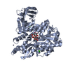
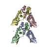
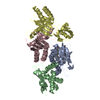
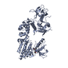
| Title | Crystal structure of full-length sperm receptor ZP3 at 2.6 A resolution | |||||||||
 Components Components | (Zona pellucida 3 ) x 2 ) x 2 | |||||||||
 Keywords Keywords | CELL ADHESION / FERTILIZATION / OOCYTE / EGG COAT / ZONA PELLUCIDA / VITELLINE ENVELOPE / ZP DOMAIN / ZP MODULE / EGG-SPERM INTERACTION / SPECIES-SPECIFIC GAMETE RECOGNITION / SPECIATION / BIODIVERSITY / INFERTILITY / EXTRACELLULAR MATRIX / IMMUNOGLOBULIN-LIKE FOLD / GLYCOPROTEIN / RECEPTOR / SECRETED / TRANSMEMBRANE / O-LINKED CARBOHYDRATE / T-ANTIGEN / CORE-1 / EXTERNAL HYDROPHOBIC PATCH / EHP / INTERNAL HYDROPHOBIC PATCH / IHP / SPERM-COMBINING SITE | |||||||||
| Function / homology |  Function and homology information Function and homology informationegg coat formation / structural constituent of egg coat / acrosin binding / positive regulation of acrosome reaction / binding of sperm to zona pellucida / response to testosterone / extracellular matrix / response to progesterone / apical part of cell / extracellular space ...egg coat formation / structural constituent of egg coat / acrosin binding / positive regulation of acrosome reaction / binding of sperm to zona pellucida / response to testosterone / extracellular matrix / response to progesterone / apical part of cell / extracellular space / identical protein binding / plasma membraneSimilarity search - Function | |||||||||
| Biological species |  Gallus gallus (chicken) Gallus gallus (chicken) | |||||||||
| Method | X-RAY DIFFRACTION / SYNCHROTRON / MOLECULAR REPLACEMENT / Resolution: 2.6 Å | |||||||||
 Authors Authors | Monne, M. / Jovine, L. | |||||||||
 Citation Citation | Journal: Cell / Year: 2010 Title: Insights into egg coat assembly and egg-sperm interaction from the X-ray structure of full-length ZP3. Authors: Ling Han / Magnus Monné / Hiroki Okumura / Thomas Schwend / Amy L Cherry / David Flot / Tsukasa Matsuda / Luca Jovine /  Abstract: ZP3, a major component of the zona pellucida (ZP) matrix coating mammalian eggs, is essential for fertilization by acting as sperm receptor. By retaining a propeptide that contains a polymerization- ...ZP3, a major component of the zona pellucida (ZP) matrix coating mammalian eggs, is essential for fertilization by acting as sperm receptor. By retaining a propeptide that contains a polymerization-blocking external hydrophobic patch (EHP), we determined the crystal structure of an avian homolog of ZP3 at 2.0 Å resolution. The structure unveils the fold of a complete ZP domain module in a homodimeric arrangement required for secretion and reveals how EHP prevents premature incorporation of ZP3 into the ZP. This suggests mechanisms underlying polymerization and how local structural differences, reflected by alternative disulfide patterns, control the specificity of ZP subunit interaction. Close relative positioning of a conserved O-glycan important for sperm binding and the hypervariable, positively selected C-terminal region of ZP3 suggests a concerted role in the regulation of species-restricted gamete recognition. Alternative conformations of the area around the O-glycan indicate how sperm binding could trigger downstream events via intramolecular signaling. #1: Journal: Cell(Cambridge,Mass.) / Year: 1980 Title: Mammalian Sperm-Egg Interaction: Identification of a Glycoprotein in Mouse Egg Zonae Pellucidae Possessing Receptor Activity for Sperm Authors: Bleil, J.D. / Wassarman, P.M. #2: Journal: FEBS Lett. / Year: 1992 Title: A Large Domain Common to Sperm Receptors (Zp2 and Zp3) and Tgf-Beta Type III Receptor Authors: Bork, P. / Sander, C. #3: Journal: Biol. Reprod. / Year: 1998 Title: The chicken homologue of zona pellucida protein-3 is synthesized by granulosa cells Authors: Waclawek, M. / Foisner, R. / Nimpf, J. / Schneider, W.J. #4: Journal: Eur.J.Biochem. / Year: 1999 Title: A 42-kDa Glycoprotein from Chicken Egg-Envelope, an Avian Homolog of the Zpc Family Glycoproteins in Mammalian Zona Pellucida. Its First Identification, Cdna Cloning and Granulosa Cell-Specific Expression. Authors: Takeuchi, Y. / Nishimura, K. / Aoki, N. / Adachi, T. / Sato, C. / Kitajima, T. / Matsuda, T. #5: Journal: Nat Cell Biol / Year: 2002 Title: The ZP domain is a conserved module for polymerization of extracellular proteins. Authors: Luca Jovine / Huayu Qi / Zev Williams / Eveline Litscher / Paul M Wassarman /  Abstract: Many eukaryotic extracellular proteins share a sequence of unknown function, called the zona pellucida (ZP) domain. Among these proteins are the mammalian sperm receptors ZP2 and ZP3, non-mammalian ...Many eukaryotic extracellular proteins share a sequence of unknown function, called the zona pellucida (ZP) domain. Among these proteins are the mammalian sperm receptors ZP2 and ZP3, non-mammalian egg coat proteins, Tamm-Horsfall protein (THP), glycoprotein-2 (GP-2), alpha- and beta-tectorins, transforming growth factor (TGF)-beta receptor III and endoglin, DMBT-1 (deleted in malignant brain tumour-1), NompA (no-mechanoreceptor-potential-A), Dumpy and cuticlin-1 (refs 1,2). Here, we report that the ZP domain of ZP2, ZP3 and THP is responsible for polymerization of these proteins into filaments of similar supramolecular structure. Most ZP domain proteins are synthesized as precursors with carboxy-terminal transmembrane domains or glycosyl phosphatidylinositol (GPI) anchors. Our results demonstrate that the C-terminal transmembrane domain and short cytoplasmic tail of ZP2 and ZP3 are not required for secretion, but are essential for assembly. Finally, we suggest a molecular basis for dominant human hearing disorders caused by point mutations within the ZP domain of alpha-tectorin. #6: Journal: Proc Natl Acad Sci U S A / Year: 2004 Title: A duplicated motif controls assembly of zona pellucida domain proteins. Authors: Luca Jovine / Huayu Qi / Zev Williams / Eveline S Litscher / Paul M Wassarman / Abstract: Many secreted eukaryotic glycoproteins that play fundamental roles in development, hearing, immunity, and cancer polymerize into filaments and extracellular matrices through zona pellucida (ZP) ...Many secreted eukaryotic glycoproteins that play fundamental roles in development, hearing, immunity, and cancer polymerize into filaments and extracellular matrices through zona pellucida (ZP) domains. ZP domain proteins are synthesized as precursors containing C-terminal propeptides that are cleaved at conserved sites. However, the consequences of this processing and the mechanism by which nascent proteins assemble are unclear. By microinjection of mutated DNA constructs into growing oocytes and mammalian cell transfection, we have identified a conserved duplicated motif [EHP (external hydrophobic patch)/IHP (internal hydrophobic patch)] regulating the assembly of mouse ZP proteins. Whereas the transmembrane domain (TMD) of ZP3 can be functionally replaced by an unrelated TMD, mutations in either EHP or IHP do not hinder secretion of full-length ZP3 but completely abolish its assembly. Because mutants truncated before the TMD are not processed, we conclude that the conserved TMD of mammalian ZP proteins does not engage them in specific interactions but is essential for C-terminal processing. Cleavage of ZP precursors results in loss of the EHP, thereby activating secreted polypeptides to assemble by using the IHP within the ZP domain. Taken together, these findings suggest a general mechanism for assembly of ZP domain proteins. #7: Journal: Annu Rev Biochem / Year: 2005 Title: Zona pellucida domain proteins. Authors: Luca Jovine / Costel C Darie / Eveline S Litscher / Paul M Wassarman / Abstract: Many eukaryotic proteins share a sequence designated as the zona pellucida (ZP) domain. This structural element, present in extracellular proteins from a wide variety of organisms, from nematodes to ...Many eukaryotic proteins share a sequence designated as the zona pellucida (ZP) domain. This structural element, present in extracellular proteins from a wide variety of organisms, from nematodes to mammals, consists of approximately 260 amino acids with eight conserved cysteine (Cys) residues and is located close to the C terminus of the polypeptide. ZP domain proteins are often glycosylated, modular structures consisting of multiple types of domains. Predictions can be made about some of the structural features of the ZP domain and ZP domain proteins. The functions of ZP domain proteins vary tremendously, from serving as structural components of egg coats, appendicularian mucous houses, and nematode dauer larvae, to serving as mechanotransducers in flies and receptors in mammals and nonmammals. Generally, ZP domain proteins are present in filaments and/or matrices, which is consistent with the role of the domain in protein polymerization. A general mechanism for assembly of ZP domain proteins has been presented. It is likely that the ZP domain plays a common role despite its presence in proteins of widely diverse functions. #8: Journal: Nature / Year: 2008Title: Crystal Structure of the Zp-N Domain of Zp3 Reveals the Core Fold of Animal Egg Coats Authors: Monne, M. / Han, L. / Schwend, T. / Burendahl, S. / Jovine, L. | |||||||||
| History |
| |||||||||
| Remark 650 | HELIX DETERMINATION METHOD: AUTHOR DETERMINED | |||||||||
| Remark 700 | SHEET DETERMINATION METHOD: AUTHOR DETERMINED |
- Structure visualization
| Structure viewer | Molecule: MolmilJmol/JSmol |
|---|
- Downloads & links
-Download
| PDBx/mmCIF format | 3nk3.cif.gz | 346.2 KB | Display | PDBx/mmCIF format |
|---|---|---|---|---|
| PDB format | pdb3nk3.ent.gz | 288.5 KB | Display | PDB format |
| PDBx/mmJSON format | 3nk3.json.gz | Tree view | PDBx/mmJSON format | |
| Others |  Other downloads Other downloads |
-Validation report
| Arichive directory | https://data.pdbj.org/pub/pdb/validation_reports/nk/3nk3ftp://data.pdbj.org/pub/pdb/validation_reports/nk/3nk3 | HTTPS FTP |
|---|
-Related structure data
| Related structure data | 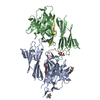 3nk4C 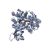 3d4gS S: Starting model for refinement C: citing same article ( |
|---|---|
| Similar structure data |


-Links
 PDBj
PDBj


- Assembly
| Deposited unit | 
| ||||||||||||||||||||||||||||||||||||||||||||||||||||||||||||||||||||||||||||||||||||||||||||||||||
|---|---|---|---|---|---|---|---|---|---|---|---|---|---|---|---|---|---|---|---|---|---|---|---|---|---|---|---|---|---|---|---|---|---|---|---|---|---|---|---|---|---|---|---|---|---|---|---|---|---|---|---|---|---|---|---|---|---|---|---|---|---|---|---|---|---|---|---|---|---|---|---|---|---|---|---|---|---|---|---|---|---|---|---|---|---|---|---|---|---|---|---|---|---|---|---|---|---|---|---|
| 1 |
| ||||||||||||||||||||||||||||||||||||||||||||||||||||||||||||||||||||||||||||||||||||||||||||||||||
| Unit cell |
| ||||||||||||||||||||||||||||||||||||||||||||||||||||||||||||||||||||||||||||||||||||||||||||||||||
| Noncrystallographic symmetry (NCS) | NCS domain:
NCS domain segments:
|