 Movie
Movie Controller


+ Open data

- Basic information
| Entry | Database: PDB / ID: 3ngy | ||||||
|---|---|---|---|---|---|---|---|
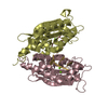
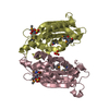
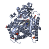
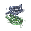
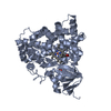
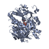
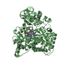
| Title | Crystal structure of RNase T (E92G mutant) | ||||||
 Components Components |
| ||||||
 Keywords Keywords |  HYDROLASE / exoribonuclease / RNA processing / RNA maturation / protein-DNA interactions / exo-nuclease HYDROLASE / exoribonuclease / RNA processing / RNA maturation / protein-DNA interactions / exo-nuclease | ||||||
| Function / homology |  Function and homology information Function and homology informationrRNA 3'-end processing / tRNA 3'-end processing / regulatory ncRNA 3'-end processing / single-stranded DNA 3'-5' DNA exonuclease activity / DNA replication proofreading / 3'-5' exonuclease activity / Hydrolases; Acting on ester bonds; Exoribonucleases producing 5'-phosphomonoesters / 3'-5'-RNA exonuclease activity / nucleic acid binding / DNA damage response ...rRNA 3'-end processing / tRNA 3'-end processing / regulatory ncRNA 3'-end processing / single-stranded DNA 3'-5' DNA exonuclease activity / DNA replication proofreading / 3'-5' exonuclease activity / Hydrolases; Acting on ester bonds; Exoribonucleases producing 5'-phosphomonoesters / 3'-5'-RNA exonuclease activity / nucleic acid binding / DNA damage response / magnesium ion binding / protein homodimerization activity / identical protein binding / cytosolSimilarity search - Function | ||||||
| Biological species |  Escherichia coli (E. coli) Escherichia coli (E. coli) | ||||||
| Method | X-RAY DIFFRACTION / SYNCHROTRON / MOLECULAR REPLACEMENT / Resolution: 2.204 Å | ||||||
 Authors Authors | Hsiao, Y.-Y. / Yuan, H.S. | ||||||
 Citation Citation | Journal: Nat.Chem.Biol. / Year: 2011 Title: Structural basis for RNA trimming by RNase T in stable RNA 3'-end maturation Authors: Hsiao, Y.-Y. / Yang, C.-C. / Lin, C.L. / Lin, J.L.J. / Duh, Y. / Yuan, H.S. | ||||||
| History |
|
- Structure visualization
| Structure viewer | Molecule: MolmilJmol/JSmol |
|---|
- Downloads & links
-Download
| PDBx/mmCIF format | 3ngy.cif.gz | 171.6 KB | Display | PDBx/mmCIF format |
|---|---|---|---|---|
| PDB format | pdb3ngy.ent.gz | 134.7 KB | Display | PDB format |
| PDBx/mmJSON format | 3ngy.json.gz | Tree view | PDBx/mmJSON format | |
| Others |  Other downloads Other downloads |
-Validation report
| Arichive directory | https://data.pdbj.org/pub/pdb/validation_reports/ng/3ngyftp://data.pdbj.org/pub/pdb/validation_reports/ng/3ngy | HTTPS FTP |
|---|
-Related structure data
| Related structure data | 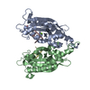 3ngzC 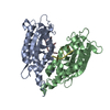 3nh0C 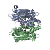 3nh1C 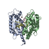 3nh2C 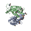 2f96S C: citing same article ( S: Starting model for refinement |
|---|---|
| Similar structure data |


-Links
 PDBj
PDBj
- Assembly
| Deposited unit | 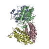
| ||||||||
|---|---|---|---|---|---|---|---|---|---|
| 1 | 
| ||||||||
| 2 | 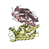
| ||||||||
| 3 | 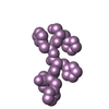
| ||||||||
| Unit cell |
|
-Components
| #1: Protein | / RNase T / Exoribonuclease T Mass: 25646.973 Da / Num. of mol.: 4 / Mutation: E92G Source method: isolated from a genetically manipulated source Source: (gene. exp.) Escherichia coli (E. coli) / Strain: JM109 / ATCC 53323 / Gene: rnt / Plasmid: pET28a / Production host: Escherichia coli (E. coli) / Strain (production host): RIPLReferences: UniProt: P30014, Hydrolases; Acting on ester bonds; Exoribonucleases producing 5'-phosphomonoesters#2: Protein/peptide | | Mass: 795.827 Da / Num. of mol.: 1 Source method: isolated from a genetically manipulated source Source: (gene. exp.) Escherichia coli (E. coli) / Strain: JM109 / ATCC 53323 / Gene: rnt / Plasmid: pET28a / Production host: Escherichia coli (E. coli) / Strain (production host): RIPL#3: Chemical |   Mass: 58.933 Da / Num. of mol.: 3 / Source method: obtained synthetically / Formula: Co Mass: 58.933 Da / Num. of mol.: 3 / Source method: obtained synthetically / Formula: Co#4: Water | ChemComp-HOH / | Water Mass: 18.015 Da / Num. of mol.: 373 / Source method: isolated from a natural source / Formula: H2O Mass: 18.015 Da / Num. of mol.: 373 / Source method: isolated from a natural source / Formula: H2OSequence details | CHAIN E IS THE N-TERMINAL HIS-TAG SEQUENCE OF ONE OF THE FOUR RNASE T SUBUNITS (CHAINS A-D). | |
|---|
-Experimental details
-Experiment
| Experiment | Method: X-RAY DIFFRACTION / Number of used crystals: 1 |
|---|
- Sample preparation
| Crystal | Density Matthews: 1.86 Å3/Da / Density % sol: 34 % |
|---|---|
| Crystal grow | Temperature: 298 K / Method: vapor diffusion, hanging drop / pH: 7 Details: 1.0M ammonium citrate tribase pH 7.0, 0.1M BIS-TRIS propane pH 7.0, 10mM ErCl3-6H2O, VAPOR DIFFUSION, HANGING DROP, temperature 298K |
-Data collection
| Diffraction | Mean temperature: 100 K |
|---|---|
| Diffraction source | Source: SYNCHROTRON / Site: NSRRC  / Beamline: BL13B1 / Wavelength: 0.999 Å / Beamline: BL13B1 / Wavelength: 0.999 Å |
| Detector | Type: ADSC QUANTUM 315 / Detector: CCD / Date: Nov 21, 2008 |
| Radiation | Monochromator: SAGITALLY FOCUSED Si(111) / Protocol: SINGLE WAVELENGTH / Monochromatic (M) / Laue (L): M / Scattering type: x-ray |
| Radiation wavelength | Wavelength: 0.999 Å / Relative weight: 1 |
| Reflection | Resolution: 2.2→30 Å / Num. all: 39447 / Num. obs: 39447 / % possible obs: 98.5 % / Observed criterion σ(F): 0 / Observed criterion σ(I): 0 / Redundancy: 2.5 % / Rsym value: 0.058 / Net I/σ(I): 23.9 |
| Reflection shell | Resolution: 2.2→2.28 Å / Redundancy: 2.3 % / Rmerge(I) obs: 0.3 / Mean I/σ(I) obs: 4.9 / Num. unique all: 3547 / Rsym value: 0.3 / % possible all: 90 |
- Processing
| Software |
| |||||||||||||||||||||||||||||||||||||||||||||||||||||||||||||||||||||||||||||||||||||||||||||||||||||||||
|---|---|---|---|---|---|---|---|---|---|---|---|---|---|---|---|---|---|---|---|---|---|---|---|---|---|---|---|---|---|---|---|---|---|---|---|---|---|---|---|---|---|---|---|---|---|---|---|---|---|---|---|---|---|---|---|---|---|---|---|---|---|---|---|---|---|---|---|---|---|---|---|---|---|---|---|---|---|---|---|---|---|---|---|---|---|---|---|---|---|---|---|---|---|---|---|---|---|---|---|---|---|---|---|---|---|---|
| Refinement | Method to determine structure: MOLECULAR REPLACEMENT Starting model: PDB 2F96 Resolution: 2.204→27.608 Å / Occupancy max: 1 / Occupancy min: 1 / FOM work R set: 0.8546 / SU ML: 0.25 / Cross valid method: THROUGHOUT / σ(F): 1.35 / Phase error: 21.19 / Stereochemistry target values: ML
| |||||||||||||||||||||||||||||||||||||||||||||||||||||||||||||||||||||||||||||||||||||||||||||||||||||||||
| Solvent computation | Shrinkage radii: 0.9 Å / VDW probe radii: 1.11 Å / Solvent model: FLAT BULK SOLVENT MODEL / Bsol: 41.096 Å2 / ksol: 0.38 e/Å3 | |||||||||||||||||||||||||||||||||||||||||||||||||||||||||||||||||||||||||||||||||||||||||||||||||||||||||
| Displacement parameters | Biso max: 84.85 Å2 / Biso mean: 27.8473 Å2 / Biso min: 9.47 Å2
| |||||||||||||||||||||||||||||||||||||||||||||||||||||||||||||||||||||||||||||||||||||||||||||||||||||||||
| Refinement step | Cycle: LAST / Resolution: 2.204→27.608 Å
| |||||||||||||||||||||||||||||||||||||||||||||||||||||||||||||||||||||||||||||||||||||||||||||||||||||||||
| Refine LS restraints |
| |||||||||||||||||||||||||||||||||||||||||||||||||||||||||||||||||||||||||||||||||||||||||||||||||||||||||
| LS refinement shell |
|
