 Movie
Movie Controller

[English] 日本語
 Yorodumi
- PDB-3n90: The 1.7 Angstrom resolution crystal structure of AT2G44920, a pen... -
+ Open data

- Basic information
| Entry | Database: PDB / ID: 3n90 | ||||||
|---|---|---|---|---|---|---|---|
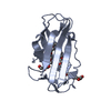
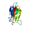
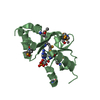
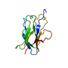
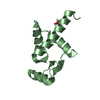
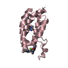
| Title | The 1.7 Angstrom resolution crystal structure of AT2G44920, a pentapeptide repeat protein from Arabidopsis thaliana thylakoid lumen. | ||||||
 Components Components | Thylakoid lumenal 15 kDa protein 1, chloroplastic | ||||||
 Keywords Keywords | UNKNOWN FUNCTION / right-handed quadrilateral beta helix /  pentapeptide repeat protein / repeated-five residue fold / chloroplast lumen pentapeptide repeat protein / repeated-five residue fold / chloroplast lumen | ||||||
| Function / homology |  Function and homology informationthylakoid lumen / chloroplast thylakoid / thylakoid / chloroplast thylakoid lumen / chloroplast thylakoid membrane / chloroplast / cytosol Function and homology informationthylakoid lumen / chloroplast thylakoid / thylakoid / chloroplast thylakoid lumen / chloroplast thylakoid membrane / chloroplast / cytosolSimilarity search - Function | ||||||
| Biological species |  Arabidopsis thaliana (thale cress) Arabidopsis thaliana (thale cress) | ||||||
| Method | X-RAY DIFFRACTION / SYNCHROTRON / SAD / Resolution: 1.7 Å | ||||||
 Authors Authors | Ni, S. / Mckgookey, M. / Tinch, S.L. / Jones, A.N. / Jayaraman, S. / Kennedy, M.A. | ||||||
 Citation Citation | Journal: To be Published Title: The 1.7 Angstrom resolution crystal structure of AT2G44920, a pentapeptide repeat protein from Arabidopsis thaliana thylakoid lumen. Authors: Ni, S. / Mckgookey, M. / Tinch, S.L. / Jones, A.N. / Jayaraman, S. / Kennedy, M.A. | ||||||
| History |
|
- Structure visualization
| Structure viewer | Molecule: MolmilJmol/JSmol |
|---|
- Downloads & links
-Download
| PDBx/mmCIF format | 3n90.cif.gz | 37.6 KB | Display | PDBx/mmCIF format |
|---|---|---|---|---|
| PDB format | pdb3n90.ent.gz | 28.9 KB | Display | PDB format |
| PDBx/mmJSON format | 3n90.json.gz | Tree view | PDBx/mmJSON format | |
| Others |  Other downloads Other downloads |
-Validation report
| Arichive directory | https://data.pdbj.org/pub/pdb/validation_reports/n9/3n90ftp://data.pdbj.org/pub/pdb/validation_reports/n9/3n90 | HTTPS FTP |
|---|
-Related structure data
| Similar structure data |
|---|


-Links
 PDBj
PDBj- Assembly
| Deposited unit | 
| ||||||||
|---|---|---|---|---|---|---|---|---|---|
| 1 |
| ||||||||
| Unit cell |
|
-Components
| #1: Protein | Mass: 16251.973 Da / Num. of mol.: 1 / Fragment: UNP residues 81-224 / Mutation: V173M, T174M Source method: isolated from a genetically manipulated source Details: T7 / Source: (gene. exp.) Arabidopsis thaliana (thale cress) / Gene: At2g44920, T13E15.7 / Plasmid: pET28b / Production host:  Escherichia coli (E. coli) / Strain (production host): BL21DE3 / References: UniProt: O22160 Escherichia coli (E. coli) / Strain (production host): BL21DE3 / References: UniProt: O22160 |
|---|---|
| #2: Chemical | ChemComp-SO4 / Sulfate  Mass: 96.063 Da / Num. of mol.: 1 / Source method: obtained synthetically / Formula: SO4 Mass: 96.063 Da / Num. of mol.: 1 / Source method: obtained synthetically / Formula: SO4 |
| #3: Water | ChemComp-HOH / Water Mass: 18.015 Da / Num. of mol.: 103 / Source method: isolated from a natural source / Formula: H2O Mass: 18.015 Da / Num. of mol.: 103 / Source method: isolated from a natural source / Formula: H2O |
-Experimental details
-Experiment
| Experiment | Method: X-RAY DIFFRACTION / Number of used crystals: 1 |
|---|
- Sample preparation
| Crystal | Density Matthews: 2.1 Å3/Da / Density % sol: 40 % |
|---|---|
| Crystal grow | Temperature: 293 K / Method: vapor diffusion, hanging drop / pH: 5.2 Details: well buffer: 2.5M ammonium sulfate, 0.5M sodium acetate; Sample buffer: 0.25M sodium chloride, 10% glycerol, 0.02M Tris, pH 7.8 protein concentration=15mg/mL, VAPOR DIFFUSION, HANGING DROP, temperature 293K |
-Data collection
| Diffraction | Mean temperature: 100 K |
|---|---|
| Diffraction source | Source: SYNCHROTRON / Site: APS  / Beamline: 24-ID-E / Wavelength: 0.979 Å / Beamline: 24-ID-E / Wavelength: 0.979 Å |
| Detector | Type: ADSC QUANTUM 315 / Detector: CCD / Date: Dec 11, 2009 Details: Cryogenically-cooled single crystal Si(111) side bounce monochromator. Optional Si(311) to achive 13.474 keV. Vertical and horizantal focussing mirrors in Kirkpatrick-Baez geometry. |
| Radiation | Monochromator: Cryogenically-cooled single crystal Si(111) side bounce monochromator. Optional Si(311) to achive 13.474 keV. Protocol: SINGLE WAVELENGTH / Monochromatic (M) / Laue (L): M / Scattering type: x-ray |
| Radiation wavelength | Wavelength: 0.979 Å / Relative weight: 1 |
| Reflection | Resolution: 1.7→50 Å / Num. all: 14963 / Num. obs: 14963 / % possible obs: 99.2 % / Observed criterion σ(F): 2 / Redundancy: 1.87 % / Biso Wilson estimate: 19.93 Å2 / Rsym value: 0.081 / Net I/σ(I): 40.84 |
| Reflection shell | Resolution: 1.7→1.79 Å / Redundancy: 1.92 % / Mean I/σ(I) obs: 18.9 / Num. unique all: 2143 / Rsym value: 0.066 / % possible all: 100 |
- Processing
| Software |
| ||||||||||||||||||||||||||||||||||||||||||||||||||||||||||||||||||||||||||||||||||||||||||||||||||||||||||||||||||||||||||||||||||||||||||||||||||||||||||||||||||||||||||
|---|---|---|---|---|---|---|---|---|---|---|---|---|---|---|---|---|---|---|---|---|---|---|---|---|---|---|---|---|---|---|---|---|---|---|---|---|---|---|---|---|---|---|---|---|---|---|---|---|---|---|---|---|---|---|---|---|---|---|---|---|---|---|---|---|---|---|---|---|---|---|---|---|---|---|---|---|---|---|---|---|---|---|---|---|---|---|---|---|---|---|---|---|---|---|---|---|---|---|---|---|---|---|---|---|---|---|---|---|---|---|---|---|---|---|---|---|---|---|---|---|---|---|---|---|---|---|---|---|---|---|---|---|---|---|---|---|---|---|---|---|---|---|---|---|---|---|---|---|---|---|---|---|---|---|---|---|---|---|---|---|---|---|---|---|---|---|---|---|---|---|---|
| Refinement | Method to determine structure: SAD / Resolution: 1.7→50 Å / Cor.coef. Fo:Fc: 0.934 / Cor.coef. Fo:Fc free: 0.925 / SU B: 2.662 / SU ML: 0.088 / Cross valid method: THROUGHOUT / ESU R Free: 0.129 / Stereochemistry target values: MAXIMUM LIKELIHOOD / Details: HYDROGENS HAVE BEEN ADDED IN THE RIDING POSITIONS
| ||||||||||||||||||||||||||||||||||||||||||||||||||||||||||||||||||||||||||||||||||||||||||||||||||||||||||||||||||||||||||||||||||||||||||||||||||||||||||||||||||||||||||
| Solvent computation | Ion probe radii: 0.8 Å / Shrinkage radii: 0.8 Å / VDW probe radii: 1.4 Å / Solvent model: MASK | ||||||||||||||||||||||||||||||||||||||||||||||||||||||||||||||||||||||||||||||||||||||||||||||||||||||||||||||||||||||||||||||||||||||||||||||||||||||||||||||||||||||||||
| Displacement parameters | Biso mean: 20.296 Å2
| ||||||||||||||||||||||||||||||||||||||||||||||||||||||||||||||||||||||||||||||||||||||||||||||||||||||||||||||||||||||||||||||||||||||||||||||||||||||||||||||||||||||||||
| Refinement step | Cycle: LAST / Resolution: 1.7→50 Å
| ||||||||||||||||||||||||||||||||||||||||||||||||||||||||||||||||||||||||||||||||||||||||||||||||||||||||||||||||||||||||||||||||||||||||||||||||||||||||||||||||||||||||||
| Refine LS restraints |
| ||||||||||||||||||||||||||||||||||||||||||||||||||||||||||||||||||||||||||||||||||||||||||||||||||||||||||||||||||||||||||||||||||||||||||||||||||||||||||||||||||||||||||
| LS refinement shell | Resolution: 1.7→1.774 Å / Total num. of bins used: 20
|
