 Movie
Movie Controller

[English] 日本語
 Yorodumi
- PDB-3n7q: Crystal structure of human mitochondrial mTERF fragment (aa 99-39... -
+ Open data

- Basic information
| Entry | Database: PDB / ID: 3n7q | ||||||
|---|---|---|---|---|---|---|---|
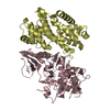
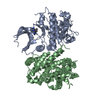
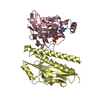
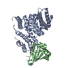
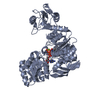
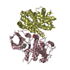
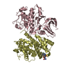
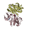
| Title | Crystal structure of human mitochondrial mTERF fragment (aa 99-399) in complex with a 12-mer DNA encompassing the tRNALeu(UUR) binding sequence | ||||||
 Components Components |
| ||||||
 Keywords Keywords |  TRANSCRIPTION / REPLICATION/DNA / Mitochondrial transcription termination factor-DNA complex / mitochondrial replication pausing-DNA complex / left-handed helical tandem repeat / Protein-DNA complex / REPLICATION-DNA complex TRANSCRIPTION / REPLICATION/DNA / Mitochondrial transcription termination factor-DNA complex / mitochondrial replication pausing-DNA complex / left-handed helical tandem repeat / Protein-DNA complex / REPLICATION-DNA complex | ||||||
| Function / homology |  Function and homology information Function and homology informationtermination of mitochondrial transcription / Mitochondrial transcription termination / DNA geometric change / mitochondrial nucleoid / DNA-templated transcription termination / Transcriptional activation of mitochondrial biogenesis / double-stranded DNA binding / nucleic acid binding / mitochondrial matrix / regulation of DNA-templated transcription ...termination of mitochondrial transcription / Mitochondrial transcription termination / DNA geometric change / mitochondrial nucleoid / DNA-templated transcription termination / Transcriptional activation of mitochondrial biogenesis / double-stranded DNA binding / nucleic acid binding / mitochondrial matrix / regulation of DNA-templated transcription / mitochondrion / RNA bindingSimilarity search - Function | ||||||
| Biological species |  Homo sapiens (human) Homo sapiens (human) | ||||||
| Method | X-RAY DIFFRACTION / SYNCHROTRON / MOLECULAR REPLACEMENT / Resolution: 2.4 Å | ||||||
 Authors Authors | Jimenez-Menendez, N. / Fernandez-Millan, P. / Rubio-Cosials, A. / Arnan, C. / Montoya, J. / Jacobs, H.T. / Bernado, P. / Coll, M. / Uson, I. / Sola, M. | ||||||
 Citation Citation | Journal: Nat.Struct.Mol.Biol. / Year: 2010 Title: Human mitochondrial mTERF wraps around DNA through a left-handed superhelical tandem repeat. Authors: Jimenez-Menendez, N. / Fernandez-Millan, P. / Rubio-Cosials, A. / Arnan, C. / Montoya, J. / Jacobs, H.T. / Bernado, P. / Coll, M. / Uson, I. / Sola, M. | ||||||
| History |
|
- Structure visualization
| Structure viewer | Molecule: MolmilJmol/JSmol |
|---|
- Downloads & links
-Download
| PDBx/mmCIF format | 3n7q.cif.gz | 88 KB | Display | PDBx/mmCIF format |
|---|---|---|---|---|
| PDB format | pdb3n7q.ent.gz | 63.9 KB | Display | PDB format |
| PDBx/mmJSON format | 3n7q.json.gz | Tree view | PDBx/mmJSON format | |
| Others |  Other downloads Other downloads |
-Validation report
| Arichive directory | https://data.pdbj.org/pub/pdb/validation_reports/n7/3n7qftp://data.pdbj.org/pub/pdb/validation_reports/n7/3n7q | HTTPS FTP |
|---|
-Related structure data


-Links
 PDBj
PDBj


- Assembly
| Deposited unit | 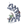
| ||||||||
|---|---|---|---|---|---|---|---|---|---|
| 1 |
| ||||||||
| Unit cell |
|
-Components
| #1: Protein | Mass: 35706.324 Da / Num. of mol.: 1 / Fragment: UNP residues 99-399 Source method: isolated from a genetically manipulated source Source: (gene. exp.) Homo sapiens (human) / Gene: MTERF / Plasmid: pET28a / Production host:  Escherichia coli (E. coli) / Strain (production host): BL21-DE3 / References: UniProt: Q99551 Escherichia coli (E. coli) / Strain (production host): BL21-DE3 / References: UniProt: Q99551 |
|---|---|
| #2: DNA chain | Mass: 3713.418 Da / Num. of mol.: 1 / Source method: obtained synthetically |
| #3: DNA chain | Mass: 3615.342 Da / Num. of mol.: 1 / Source method: obtained synthetically |
| #4: Chemical | ChemComp-CIT / Citric acid  Mass: 192.124 Da / Num. of mol.: 1 / Source method: obtained synthetically / Formula: C6H8O7 Mass: 192.124 Da / Num. of mol.: 1 / Source method: obtained synthetically / Formula: C6H8O7 |
| #5: Water | ChemComp-HOH / Water Mass: 18.015 Da / Num. of mol.: 32 / Source method: isolated from a natural source / Formula: H2O Mass: 18.015 Da / Num. of mol.: 32 / Source method: isolated from a natural source / Formula: H2O |
-Experimental details
-Experiment
| Experiment | Method: X-RAY DIFFRACTION / Number of used crystals: 1 |
|---|
- Sample preparation
| Crystal | Density Matthews: 2.65 Å3/Da / Density % sol: 53.56 % |
|---|---|
| Crystal grow | Method: vapor diffusion / Details: VAPOR DIFFUSION |
-Data collection
| Diffraction | Mean temperature: 100 K |
|---|---|
| Diffraction source | Source: SYNCHROTRON / Site: ESRF  / Beamline: ID29 / Beamline: ID29 |
| Detector | Detector: CCD / Date: Nov 5, 2008 |
| Radiation | Protocol: SINGLE WAVELENGTH / Monochromatic (M) / Laue (L): M / Scattering type: x-ray |
| Radiation wavelength | Relative weight: 1 |
| Reflection | Resolution: 2.4→31 Å / Num. all: 17262 / Num. obs: 17262 |
| Reflection shell | Resolution: 2.4→2.55 Å / Num. unique all: 165 |
- Processing
| Software |
| |||||||||||||||||||||||||||||||||||||||||||||||||
|---|---|---|---|---|---|---|---|---|---|---|---|---|---|---|---|---|---|---|---|---|---|---|---|---|---|---|---|---|---|---|---|---|---|---|---|---|---|---|---|---|---|---|---|---|---|---|---|---|---|---|
| Refinement | Method to determine structure: MOLECULAR REPLACEMENT / Resolution: 2.4→31 Å / SU ML: 0.39 / σ(F): 1.97 / Stereochemistry target values: ML
| |||||||||||||||||||||||||||||||||||||||||||||||||
| Solvent computation | Shrinkage radii: 0.9 Å / VDW probe radii: 1.11 Å / Solvent model: FLAT BULK SOLVENT MODEL / Bsol: 61.005 Å2 / ksol: 0.323 e/Å3 | |||||||||||||||||||||||||||||||||||||||||||||||||
| Displacement parameters |
| |||||||||||||||||||||||||||||||||||||||||||||||||
| Refinement step | Cycle: LAST / Resolution: 2.4→31 Å
| |||||||||||||||||||||||||||||||||||||||||||||||||
| Refine LS restraints |
| |||||||||||||||||||||||||||||||||||||||||||||||||
| LS refinement shell |
|
