 Movie
Movie Controller

[English] 日本語
 Yorodumi
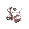
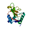
- PDB-3n7c: Crystal structure of the RAN binding domain from the nuclear pore... -
+ Open data

- Basic information
| Entry | Database: PDB / ID: 3n7c | ||||||
|---|---|---|---|---|---|---|---|
| Title | Crystal structure of the RAN binding domain from the nuclear pore complex component NUP2 from Ashbya gossypii | ||||||
 Components Components | ABR034Wp | ||||||
 Keywords Keywords |  PROTEIN TRANSPORT / Nuclear pore complex / NUP2 / Ran-binding domain / Nucleoporin / STRUCTURAL GENOMICS / PSI-2 / PROTEIN STRUCTURE INITIATIVE / NEW YORK STRUCTURAL GENOMIX RESEARCH CONSORTIUM / NYSGXRC / New York SGX Research Center for Structural Genomics PROTEIN TRANSPORT / Nuclear pore complex / NUP2 / Ran-binding domain / Nucleoporin / STRUCTURAL GENOMICS / PSI-2 / PROTEIN STRUCTURE INITIATIVE / NEW YORK STRUCTURAL GENOMIX RESEARCH CONSORTIUM / NYSGXRC / New York SGX Research Center for Structural Genomics | ||||||
| Function / homology |  Function and homology information Function and homology informationmRNA export from nucleus in response to heat stress / protein localization to nuclear inner membrane / post-transcriptional tethering of RNA polymerase II gene DNA at nuclear periphery / nuclear pore cytoplasmic filaments / nuclear pore nuclear basket / importin-alpha family protein binding / NLS-dependent protein nuclear import complex / structural constituent of nuclear pore / silent mating-type cassette heterochromatin formation / poly(A)+ mRNA export from nucleus ...mRNA export from nucleus in response to heat stress / protein localization to nuclear inner membrane / post-transcriptional tethering of RNA polymerase II gene DNA at nuclear periphery / nuclear pore cytoplasmic filaments / nuclear pore nuclear basket / importin-alpha family protein binding / NLS-dependent protein nuclear import complex / structural constituent of nuclear pore / silent mating-type cassette heterochromatin formation / poly(A)+ mRNA export from nucleus / NLS-bearing protein import into nucleus / subtelomeric heterochromatin formation / nuclear pore / protein export from nucleus / small GTPase binding / chromosome, telomeric region / cytoplasmSimilarity search - Function | ||||||
| Biological species |  Ashbya gossypii (fungus) Ashbya gossypii (fungus) | ||||||
| Method | X-RAY DIFFRACTION / SYNCHROTRON / SAD / Resolution: 2.26 Å | ||||||
 Authors Authors | Sampathkumar, P. / Manglicmot, D. / Gilmore, J. / Bain, K. / Gheyi, T. / Atwell, S. / Thompson, D.A. / Emtage, J.S. / Wasserman, S. / Sauder, J.M. ...Sampathkumar, P. / Manglicmot, D. / Gilmore, J. / Bain, K. / Gheyi, T. / Atwell, S. / Thompson, D.A. / Emtage, J.S. / Wasserman, S. / Sauder, J.M. / Burley, S.K. / New York SGX Research Center for Structural Genomics (NYSGXRC) | ||||||
 Citation Citation | Journal: To be Published Title: Crystal structure of the RAN binding domain from the nuclear pore complex component NUP2 from Ashbya gossypii Authors: Sampathkumar, P. | ||||||
| History |
|
- Structure visualization
| Structure viewer | Molecule: MolmilJmol/JSmol |
|---|
- Downloads & links
-Download
| PDBx/mmCIF format | 3n7c.cif.gz | 50.7 KB | Display | PDBx/mmCIF format |
|---|---|---|---|---|
| PDB format | pdb3n7c.ent.gz | 38.4 KB | Display | PDB format |
| PDBx/mmJSON format | 3n7c.json.gz | Tree view | PDBx/mmJSON format | |
| Others |  Other downloads Other downloads |
-Validation report
| Arichive directory | https://data.pdbj.org/pub/pdb/validation_reports/n7/3n7cftp://data.pdbj.org/pub/pdb/validation_reports/n7/3n7c | HTTPS FTP |
|---|
-Related structure data
| Similar structure data | |
|---|---|
| Other databases |


-Links
 PDBj
PDBj
- Assembly
| Deposited unit | 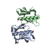
| ||||||||
|---|---|---|---|---|---|---|---|---|---|
| 1 | 
| ||||||||
| 2 | 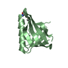
| ||||||||
| Unit cell |
|
-Components
| #1: Protein | Mass: 14820.566 Da / Num. of mol.: 2 / Fragment: sequence database residues 497-615 Source method: isolated from a genetically manipulated source Source: (gene. exp.) Ashbya gossypii (fungus) / Strain: ATCC 10895 / Gene: ABR034W, AGOS_ABR034W, NUP2 / Plasmid: BC-pSGX3(BC); modified pET26b / Production host:  Escherichia coli (E. coli) / Strain (production host): BL21(DE3)-Codon+RIL / References: UniProt: Q75DJ0 Escherichia coli (E. coli) / Strain (production host): BL21(DE3)-Codon+RIL / References: UniProt: Q75DJ0#2: Water | ChemComp-HOH / | Water Mass: 18.015 Da / Num. of mol.: 27 / Source method: isolated from a natural source / Formula: H2O Mass: 18.015 Da / Num. of mol.: 27 / Source method: isolated from a natural source / Formula: H2O |
|---|
-Experimental details
-Experiment
| Experiment | Method: X-RAY DIFFRACTION / Number of used crystals: 1 |
|---|
- Sample preparation
| Crystal | Density Matthews: 2.31 Å3/Da / Density % sol: 46.86 % |
|---|---|
| Crystal grow | Temperature: 294 K / Method: vapor diffusion, sitting drop / pH: 5.5 Details: 100mM Bis-Tris pH 5.5 + 25% PEG 3350, VAPOR DIFFUSION, SITTING DROP, temperature 294K |
-Data collection
| Diffraction | Mean temperature: 100 K |
|---|---|
| Diffraction source | Source: SYNCHROTRON / Site: APS  / Beamline: 31-ID / Wavelength: 0.97929 Å / Beamline: 31-ID / Wavelength: 0.97929 Å |
| Detector | Type: RAYONIX MX225HE / Detector: CCD / Date: Feb 10, 2010 |
| Radiation | Monochromator: diamond / Protocol: SINGLE WAVELENGTH / Monochromatic (M) / Laue (L): M / Scattering type: x-ray |
| Radiation wavelength | Wavelength: 0.97929 Å / Relative weight: 1 |
| Reflection | Resolution: 2.26→38.12 Å / Num. obs: 13396 / % possible obs: 99.7 % / Redundancy: 13.5 % / Biso Wilson estimate: 42.54 Å2 / Rsym value: 0.103 / Net I/σ(I): 15.7 |
| Reflection shell | Resolution: 2.26→2.38 Å / Redundancy: 13.7 % / Mean I/σ(I) obs: 5.3 / Num. unique all: 1925 / Rsym value: 0.58 / % possible all: 100 |
- Processing
| Software |
| |||||||||||||||||||||||||||||||||||||||||||||||||||||||||||||||||||||||||||||||||||||
|---|---|---|---|---|---|---|---|---|---|---|---|---|---|---|---|---|---|---|---|---|---|---|---|---|---|---|---|---|---|---|---|---|---|---|---|---|---|---|---|---|---|---|---|---|---|---|---|---|---|---|---|---|---|---|---|---|---|---|---|---|---|---|---|---|---|---|---|---|---|---|---|---|---|---|---|---|---|---|---|---|---|---|---|---|---|---|
| Refinement | Method to determine structure: SAD / Resolution: 2.26→21.17 Å / Cor.coef. Fo:Fc: 0.927 / Cor.coef. Fo:Fc free: 0.906 / Occupancy max: 1 / Occupancy min: 0.5 / SU B: 7.248 / SU ML: 0.183 / Cross valid method: THROUGHOUT / σ(F): 0 / ESU R: 0.276 / ESU R Free: 0.241 Stereochemistry target values: MAXIMUM LIKELIHOOD WITH PHASES Details: HYDROGENS HAVE BEEN ADDED IN THE RIDING POSITIONS U VALUES : REFINED INDIVIDUALLY
| |||||||||||||||||||||||||||||||||||||||||||||||||||||||||||||||||||||||||||||||||||||
| Solvent computation | Ion probe radii: 0.8 Å / Shrinkage radii: 0.8 Å / VDW probe radii: 1.4 Å / Solvent model: BABINET MODEL WITH MASK | |||||||||||||||||||||||||||||||||||||||||||||||||||||||||||||||||||||||||||||||||||||
| Displacement parameters | Biso max: 71.73 Å2 / Biso mean: 43.78 Å2 / Biso min: 23.93 Å2
| |||||||||||||||||||||||||||||||||||||||||||||||||||||||||||||||||||||||||||||||||||||
| Refinement step | Cycle: LAST / Resolution: 2.26→21.17 Å
| |||||||||||||||||||||||||||||||||||||||||||||||||||||||||||||||||||||||||||||||||||||
| Refine LS restraints |
| |||||||||||||||||||||||||||||||||||||||||||||||||||||||||||||||||||||||||||||||||||||
| LS refinement shell | Resolution: 2.26→2.318 Å / Total num. of bins used: 20
|
