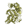
 Movie
Movie Controller


+ Open data

- Basic information
| Entry | Database: PDB / ID: 3n5g | ||||||
|---|---|---|---|---|---|---|---|
| Title | Crystal Structure of histidine-tagged human thymidylate synthase | ||||||
 Components Components | Thymidylate synthase | ||||||
 Keywords Keywords | TRANSFERASE / peptide inhibitor / protein-peptide complex / interface inhibitor | ||||||
| Function / homology |  Function and homology information Function and homology informationuracil metabolic process / response to organophosphorus / intestinal epithelial cell maturation / response to folic acid / Interconversion of nucleotide di- and triphosphates / response to vitamin A / thymidylate synthase / sequence-specific mRNA binding / cartilage development / tetrahydrofolate interconversion ...uracil metabolic process / response to organophosphorus / intestinal epithelial cell maturation / response to folic acid / Interconversion of nucleotide di- and triphosphates / response to vitamin A / thymidylate synthase / sequence-specific mRNA binding / cartilage development / tetrahydrofolate interconversion / thymidylate synthase activity / folic acid binding / dTMP biosynthetic process / dTTP biosynthetic process / DNA biosynthetic process / G1/S-Specific Transcription / developmental growth / dihydrofolate reductase activity / response to glucocorticoid / mRNA regulatory element binding translation repressor activity / response to progesterone / response to cytokine / liver regeneration / response to toxic substance / circadian rhythm / methylation / response to ethanol / mitochondrial inner membrane / negative regulation of translation / mitochondrial matrix / response to xenobiotic stimulus / protein homodimerization activity / mitochondrion / nucleus / cytosol / cytoplasmSimilarity search - Function | ||||||
| Biological species |  Homo sapiens (human) Homo sapiens (human) | ||||||
| Method | X-RAY DIFFRACTION / SYNCHROTRON / MOLECULAR REPLACEMENT / Resolution: 2.27 Å | ||||||
 Authors Authors | Pozzi, C. / Cardinale, D. / Guaitoli, G. / Tondi, D. / Luciani, R. / Myllykallio, H. / Ferrari, S. / Costi, M.P. / Mangani, S. | ||||||
 Citation Citation | Journal: Proc.Natl.Acad.Sci.USA / Year: 2011 Title: Protein-protein interface-binding peptides inhibit the cancer therapy target human thymidylate synthase. Authors: Cardinale, D. / Guaitoli, G. / Tondi, D. / Luciani, R. / Henrich, S. / Salo-Ahen, O.M. / Ferrari, S. / Marverti, G. / Guerrieri, D. / Ligabue, A. / Frassineti, C. / Pozzi, C. / Mangani, S. / ...Authors: Cardinale, D. / Guaitoli, G. / Tondi, D. / Luciani, R. / Henrich, S. / Salo-Ahen, O.M. / Ferrari, S. / Marverti, G. / Guerrieri, D. / Ligabue, A. / Frassineti, C. / Pozzi, C. / Mangani, S. / Fessas, D. / Guerrini, R. / Ponterini, G. / Wade, R.C. / Costi, M.P. | ||||||
| History |
|
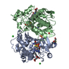
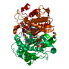
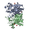
- Structure visualization
| Structure viewer | Molecule: MolmilJmol/JSmol |
|---|
- Downloads & links
-Download
| PDBx/mmCIF format | 3n5g.cif.gz | 129.6 KB | Display | PDBx/mmCIF format |
|---|---|---|---|---|
| PDB format | pdb3n5g.ent.gz | 100.5 KB | Display | PDB format |
| PDBx/mmJSON format | 3n5g.json.gz | Tree view | PDBx/mmJSON format | |
| Others |  Other downloads Other downloads |
-Validation report
| Arichive directory | https://data.pdbj.org/pub/pdb/validation_reports/n5/3n5gftp://data.pdbj.org/pub/pdb/validation_reports/n5/3n5g | HTTPS FTP |
|---|
-Related structure data
| Related structure data |  3n5eC  1ypvS C: citing same article ( S: Starting model for refinement |
|---|---|
| Similar structure data |


-Links
 PDBj
PDBj


- Assembly
| Deposited unit | 
| ||||||||
|---|---|---|---|---|---|---|---|---|---|
| 1 | 
| ||||||||
| Unit cell |
|
-Components
| #1: Protein | / TSase / TS Mass: 37382.848 Da / Num. of mol.: 1 Source method: isolated from a genetically manipulated source Source: (gene. exp.) Homo sapiens (human) / Gene: TYMS, TS, OK/SW-cl.29 / Production host:  Escherichia coli (E. coli) / References: UniProt: P04818, thymidylate synthase Escherichia coli (E. coli) / References: UniProt: P04818, thymidylate synthase | ||
|---|---|---|---|
| #2: Chemical | Sulfate  Mass: 96.063 Da / Num. of mol.: 3 / Source method: obtained synthetically / Formula: SO4 Mass: 96.063 Da / Num. of mol.: 3 / Source method: obtained synthetically / Formula: SO4#3: Water | ChemComp-HOH / | Water Mass: 18.015 Da / Num. of mol.: 149 / Source method: isolated from a natural source / Formula: H2O Mass: 18.015 Da / Num. of mol.: 149 / Source method: isolated from a natural source / Formula: H2O |
-Experimental details
-Experiment
| Experiment | Method: X-RAY DIFFRACTION / Number of used crystals: 1 |
|---|
- Sample preparation
| Crystal | Density Matthews: 2.96 Å3/Da / Density % sol: 58.51 % |
|---|---|
| Crystal grow | Temperature: 293 K / Method: vapor diffusion, sitting drop / pH: 8.3 Details: 30% saturated Ammonium Sulfate + 20mM BME + 0.1M TRIS pH8.3, VAPOR DIFFUSION, SITTING DROP, temperature 293.0K |
-Data collection
| Diffraction | Mean temperature: 100 K |
|---|---|
| Diffraction source | Source: SYNCHROTRON / Site: ESRF  / Beamline: ID14-1 / Wavelength: 0.934 Å / Beamline: ID14-1 / Wavelength: 0.934 Å |
| Detector | Type: ADSC QUANTUM 210 / Detector: CCD / Date: Apr 20, 2008 |
| Radiation | Monochromator: Diamond (111), Ge(220) / Protocol: SINGLE WAVELENGTH / Monochromatic (M) / Laue (L): M / Scattering type: x-ray |
| Radiation wavelength | Wavelength: 0.934 Å / Relative weight: 1 |
| Reflection | Resolution: 2.27→41.6 Å / Num. obs: 20905 / % possible obs: 100 % / Observed criterion σ(F): 0 / Observed criterion σ(I): 2 / Redundancy: 10.8 % / Biso Wilson estimate: 39.8 Å2 / Rmerge(I) obs: 0.07 / Rsym value: 0.07 / Net I/σ(I): 26 |
| Reflection shell | Resolution: 2.27→2.39 Å / Redundancy: 11 % / Rmerge(I) obs: 0.351 / Mean I/σ(I) obs: 7.1 / Num. unique all: 2988 / Rsym value: 0.351 / % possible all: 100 |
- Processing
| Software |
| |||||||||||||||||||||||||||||||||||||||||||||||||||||||||||||||||
|---|---|---|---|---|---|---|---|---|---|---|---|---|---|---|---|---|---|---|---|---|---|---|---|---|---|---|---|---|---|---|---|---|---|---|---|---|---|---|---|---|---|---|---|---|---|---|---|---|---|---|---|---|---|---|---|---|---|---|---|---|---|---|---|---|---|---|
| Refinement | Method to determine structure: MOLECULAR REPLACEMENT Starting model: PDB ENTRY 1YPV Resolution: 2.27→41.6 Å / Cor.coef. Fo:Fc: 0.96 / Cor.coef. Fo:Fc free: 0.942 / SU B: 8.679 / SU ML: 0.099 / Cross valid method: THROUGHOUT / σ(F): 0 / σ(I): 0 / ESU R: 0.179 / ESU R Free: 0.159 / Stereochemistry target values: MAXIMUM LIKELIHOOD
| |||||||||||||||||||||||||||||||||||||||||||||||||||||||||||||||||
| Solvent computation | Ion probe radii: 0.8 Å / Shrinkage radii: 0.8 Å / VDW probe radii: 1.4 Å / Solvent model: MASK | |||||||||||||||||||||||||||||||||||||||||||||||||||||||||||||||||
| Displacement parameters | Biso mean: 43.509 Å2
| |||||||||||||||||||||||||||||||||||||||||||||||||||||||||||||||||
| Refine analyze |
| |||||||||||||||||||||||||||||||||||||||||||||||||||||||||||||||||
| Refinement step | Cycle: LAST / Resolution: 2.27→41.6 Å
| |||||||||||||||||||||||||||||||||||||||||||||||||||||||||||||||||
| Refine LS restraints |
| |||||||||||||||||||||||||||||||||||||||||||||||||||||||||||||||||
| LS refinement shell | Resolution: 2.27→2.329 Å / Total num. of bins used: 20
| |||||||||||||||||||||||||||||||||||||||||||||||||||||||||||||||||
| Refinement TLS params. | Method: refined / Origin x: 49.3003 Å / Origin y: -7.7524 Å / Origin z: 15.8894 Å
|
