 Movie
Movie Controller

[English] 日本語
 Yorodumi
- PDB-3n3b: Ribonucleotide Reductase Dimanganese(II)-NrdF from Escherichia co... -
+ Open data

- Basic information
| Entry | Database: PDB / ID: 3n3b | ||||||
|---|---|---|---|---|---|---|---|
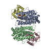
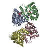
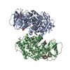
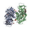
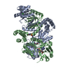
| Title | Ribonucleotide Reductase Dimanganese(II)-NrdF from Escherichia coli in Complex with Reduced NrdI with a Trapped Peroxide | ||||||
 Components Components |
| ||||||
 Keywords Keywords |  OXIDOREDUCTASE / ribonucleotide reductase / four-helix bundle / dimanganese cluster / flavoprotein / peroxide OXIDOREDUCTASE / ribonucleotide reductase / four-helix bundle / dimanganese cluster / flavoprotein / peroxide | ||||||
| Function / homology |  Function and homology information Function and homology informationribonucleoside diphosphate metabolic process / 2'-deoxyribonucleotide biosynthetic process / ribonucleoside-diphosphate reductase complex / ribonucleoside-diphosphate reductase / ribonucleoside-diphosphate reductase activity, thioredoxin disulfide as acceptor / deoxyribonucleotide biosynthetic process / protein modification process / FMN binding / manganese ion bindingSimilarity search - Function | ||||||
| Biological species |  Escherichia coli (E. coli) Escherichia coli (E. coli) | ||||||
| Method | X-RAY DIFFRACTION / SYNCHROTRON / MOLECULAR REPLACEMENT / Resolution: 2.36 Å | ||||||
 Authors Authors | Boal, A.K. / Cotruvo Jr., J.A. / Stubbe, J. / Rosenzweig, A.C. | ||||||
 Citation Citation | Journal: Science / Year: 2010 Title: Structural basis for activation of class Ib ribonucleotide reductase. Authors: Boal, A.K. / Cotruvo, J.A. / Stubbe, J. / Rosenzweig, A.C. | ||||||
| History |
|
- Structure visualization
| Structure viewer | Molecule: MolmilJmol/JSmol |
|---|
- Downloads & links
-Download
| PDBx/mmCIF format | 3n3b.cif.gz | 337 KB | Display | PDBx/mmCIF format |
|---|---|---|---|---|
| PDB format | pdb3n3b.ent.gz | 274.5 KB | Display | PDB format |
| PDBx/mmJSON format | 3n3b.json.gz | Tree view | PDBx/mmJSON format | |
| Others |  Other downloads Other downloads |
-Validation report
| Arichive directory | https://data.pdbj.org/pub/pdb/validation_reports/n3/3n3bftp://data.pdbj.org/pub/pdb/validation_reports/n3/3n3b | HTTPS FTP |
|---|
-Related structure data
| Related structure data | 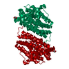 3n37C 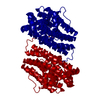 3n38C  3n39SC 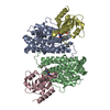 3n3aC C: citing same article ( S: Starting model for refinement |
|---|---|
| Similar structure data |

-Links
 PDBj
PDBj
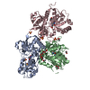
- Assembly
| Deposited unit | 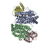
| ||||||||
|---|---|---|---|---|---|---|---|---|---|
| 1 |
| ||||||||
| Unit cell |
|
-Components
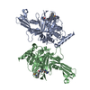
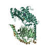
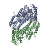
-Protein , 2 types, 4 molecules BACD
| #1: Protein | Ribonucleotide reductase / Ribonucleotide reductase 2 / R2F protein Mass: 36475.070 Da / Num. of mol.: 2 Source method: isolated from a genetically manipulated source Source: (gene. exp.) Escherichia coli (E. coli) / Strain: K12 / Gene: b2676, JW2651, nrdF, ygaD / Production host: Escherichia coli (E. coli)References: UniProt: P37146, ribonucleoside-diphosphate reductase#2: Protein | Mass: 17216.404 Da / Num. of mol.: 2 Source method: isolated from a genetically manipulated source Source: (gene. exp.) Escherichia coli (E. coli) / Strain: K12 / Gene: nrdI, ygaO, b2674, JW2649 / Production host: Escherichia coli (E. coli) / References: UniProt: P0A772 |
|---|
-Non-polymers , 4 types, 170 molecules 


| #3: Chemical | ChemComp-MN /  Mass: 54.938 Da / Num. of mol.: 4 / Source method: obtained synthetically / Formula: Mn Mass: 54.938 Da / Num. of mol.: 4 / Source method: obtained synthetically / Formula: Mn#4: Chemical | Hydrogen peroxide Mass: 34.015 Da / Num. of mol.: 2 / Source method: obtained synthetically / Formula: H2O2 Mass: 34.015 Da / Num. of mol.: 2 / Source method: obtained synthetically / Formula: H2O2#5: Chemical | Flavin mononucleotide Mass: 456.344 Da / Num. of mol.: 2 / Source method: obtained synthetically / Formula: C17H21N4O9P Mass: 456.344 Da / Num. of mol.: 2 / Source method: obtained synthetically / Formula: C17H21N4O9P#6: Water | ChemComp-HOH / | WaterMass: 18.015 Da / Num. of mol.: 162 / Source method: isolated from a natural source / Formula: H2O |
|---|
-Experimental details
-Experiment
| Experiment | Method: X-RAY DIFFRACTION / Number of used crystals: 1 |
|---|
- Sample preparation
| Crystal | Density Matthews: 2.32 Å3/Da / Density % sol: 47 % |
|---|
-Data collection
| Diffraction | Mean temperature: 113 K |
|---|---|
| Diffraction source | Source: SYNCHROTRON / Site: APS  / Beamline: 21-ID-D / Wavelength: 1.0781 Å / Beamline: 21-ID-D / Wavelength: 1.0781 Å |
| Detector | Type: MARMOSAIC 225 mm CCD / Detector: CCD / Date: Dec 21, 2009 |
| Radiation | Protocol: SINGLE WAVELENGTH / Monochromatic (M) / Laue (L): M / Scattering type: x-ray |
| Radiation wavelength | Wavelength: 1.0781 Å / Relative weight: 1 |
| Reflection | Resolution: 2.35→40.56 Å |
- Processing
| Software |
| |||||||||||||||||||||||||||||||||||||||||||||||||||||||||||||||||||||||||||||||||||||||||||||||||||||||||||||||||||||||||||||
|---|---|---|---|---|---|---|---|---|---|---|---|---|---|---|---|---|---|---|---|---|---|---|---|---|---|---|---|---|---|---|---|---|---|---|---|---|---|---|---|---|---|---|---|---|---|---|---|---|---|---|---|---|---|---|---|---|---|---|---|---|---|---|---|---|---|---|---|---|---|---|---|---|---|---|---|---|---|---|---|---|---|---|---|---|---|---|---|---|---|---|---|---|---|---|---|---|---|---|---|---|---|---|---|---|---|---|---|---|---|---|---|---|---|---|---|---|---|---|---|---|---|---|---|---|---|---|
| Refinement | Method to determine structure: MOLECULAR REPLACEMENT Starting model: 3N39 Resolution: 2.36→40.56 Å / Cor.coef. Fo:Fc: 0.917 / Cor.coef. Fo:Fc free: 0.889 / Occupancy max: 1 / Occupancy min: 0.5 / SU B: 17.302 / SU ML: 0.2 / Cross valid method: THROUGHOUT / σ(F): 0 / ESU R Free: 0.29 / Stereochemistry target values: MAXIMUM LIKELIHOOD / Details: HYDROGENS HAVE BEEN ADDED IN THE RIDING POSITIONS
| |||||||||||||||||||||||||||||||||||||||||||||||||||||||||||||||||||||||||||||||||||||||||||||||||||||||||||||||||||||||||||||
| Solvent computation | Ion probe radii: 0.8 Å / Shrinkage radii: 0.8 Å / VDW probe radii: 1.4 Å / Solvent model: MASK | |||||||||||||||||||||||||||||||||||||||||||||||||||||||||||||||||||||||||||||||||||||||||||||||||||||||||||||||||||||||||||||
| Displacement parameters | Biso max: 60.11 Å2 / Biso mean: 33.897 Å2 / Biso min: 5.96 Å2
| |||||||||||||||||||||||||||||||||||||||||||||||||||||||||||||||||||||||||||||||||||||||||||||||||||||||||||||||||||||||||||||
| Refinement step | Cycle: LAST / Resolution: 2.36→40.56 Å
| |||||||||||||||||||||||||||||||||||||||||||||||||||||||||||||||||||||||||||||||||||||||||||||||||||||||||||||||||||||||||||||
| Refine LS restraints |
| |||||||||||||||||||||||||||||||||||||||||||||||||||||||||||||||||||||||||||||||||||||||||||||||||||||||||||||||||||||||||||||
| LS refinement shell | Resolution: 2.356→2.417 Å / Total num. of bins used: 20
| |||||||||||||||||||||||||||||||||||||||||||||||||||||||||||||||||||||||||||||||||||||||||||||||||||||||||||||||||||||||||||||
| Refinement TLS params. | Method: refined / Refine-ID: X-RAY DIFFRACTION
| |||||||||||||||||||||||||||||||||||||||||||||||||||||||||||||||||||||||||||||||||||||||||||||||||||||||||||||||||||||||||||||
| Refinement TLS group |
|
