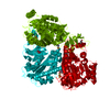
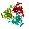
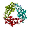
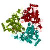
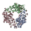
CRYSTAL PACKING ANALYSIS AND ANALYTICAL SIZE EXCLUSION CHROMATOGRAPHY COUPLED WITH STATIC LIGHT SCATTERING SUPPORT THE ASSIGNMENT OF A TRIMER AS A SIGNIFICANT OLIGOMERIC FORM IN SOLUTION.
-
Components
-
Protein , 1 types, 1 molecules A
#1: Protein
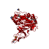
Putativearomatic-ringhydroxylatingdioxygenase / Probable Ring hydroxylating alpha subunit
Mass: 46487.980 Da / Num. of mol.: 1 Source method: isolated from a genetically manipulated source Source: (gene. exp.) Ruegeria sp. TM1040 (bacteria) / Gene: benA, TM1040_3219 / Plasmid: SpeedET / Production host: Escherichia Coli (E. coli) / Strain (production host): HK100 / References: UniProt: Q1GMC3
Mass: 18.015 Da / Num. of mol.: 469 / Source method: isolated from a natural source / Formula: H2O
-
Details
Sequence details
THIS CONSTRUCT WAS EXPRESSED WITH AN N-TERMINAL PURIFICATION TAG MGSDKIHHHHHHENLYFQG. THE TAG WAS ...THIS CONSTRUCT WAS EXPRESSED WITH AN N-TERMINAL PURIFICATION TAG MGSDKIHHHHHHENLYFQG. THE TAG WAS REMOVED WITH TEV PROTEASE LEAVING ONLY A GLYCINE (0) FOLLOWED BY THE TARGET SEQUENCE.
-
Experimental details
-
Experiment
Experiment
Method: X-RAY DIFFRACTION / Number of used crystals: 1
-
Sample preparation
Crystal
Density Matthews: 2.85 Å3/Da / Density % sol: 56.85 %
Crystal grow
Temperature: 277 K / Method: vapor diffusion, sitting drop / pH: 8.29 Details: 23.8000% polyethylene glycol monomethyl ether 2000, 0.0100M nickel (II) chloride, 0.1M TRIS pH 8.29, NANODROP', VAPOR DIFFUSION, SITTING DROP, temperature 277K
Type: MARMOSAIC 325 mm CCD / Detector: CCD / Date: Feb 10, 2010 / Details: Flat mirror (vertical focusing)
Radiation
Monochromator: Single crystal Si(111) bent monochromator (horizontal focusing) Protocol: MAD / Monochromatic (M) / Laue (L): M / Scattering type: x-ray
Radiation wavelength
ID
Wavelength (Å)
Relative weight
1
0.91837
1
2
0.97949
1
3
0.97895
1
Reflection
Resolution: 1.8→28.155 Å / Num. obs: 48265 / % possible obs: 99.3 % / Observed criterion σ(I): -3 / Redundancy: 6.31 % / Biso Wilson estimate: 22.737 Å2 / Rmerge(I) obs: 0.06 / Net I/σ(I): 14.33
Reflection shell
Resolution (Å)
Rmerge(I) obs
Mean I/σ(I) obs
Num. measured obs
Num. unique obs
% possible all
1.8-1.86
0.577
2.16
28099
8783
98.9
1.86-1.94
0.421
3
32555
10184
99.1
1.94-2.03
0.274
4.4
30765
9585
99.2
2.03-2.13
0.199
6.1
28346
8822
99.2
2.13-2.27
0.14
8.4
32011
9942
99.3
2.27-2.44
0.1
11.4
29634
9189
99.4
2.44-2.69
0.076
14.8
31247
9675
99.5
2.69-3.07
0.052
20.7
29995
9290
99.6
3.07-3.87
0.034
31.4
30874
9574
99.7
3.87-28.155
0.024
40.4
30919
9582
99.5
-
Phasing
Phasing
Method: MAD
-
Processing
Software
Name
Version
Classification
NB
REFMAC
5.6.0073
refinement
PHENIX
refinement
SHELX
phasing
MolProbity
3beta29
modelbuilding
XSCALE
datascaling
PDB_EXTRACT
3.006
dataextraction
XDS
datareduction
autoSHARP
phasing
Refinement
Method to determine structure: MAD / Resolution: 1.8→28.155 Å / Cor.coef. Fo:Fc: 0.965 / Cor.coef. Fo:Fc free: 0.951 / Occupancy max: 1 / Occupancy min: 0.3 / SU B: 3.589 / SU ML: 0.063 / Cross valid method: THROUGHOUT / σ(F): 0 / ESU R Free: 0.098 Stereochemistry target values: MAXIMUM LIKELIHOOD WITH PHASES Details: 1. HYDROGENS HAVE BEEN ADDED IN THE RIDING POSITIONS. 2. A MET-INHIBITION PROTOCOL WAS USED FOR SELENOMETHIONINE INCORPORATION DURING PROTEIN EXPRESSION. THE OCCUPANCY OF THE SE ATOMS IN THE ...Details: 1. HYDROGENS HAVE BEEN ADDED IN THE RIDING POSITIONS. 2. A MET-INHIBITION PROTOCOL WAS USED FOR SELENOMETHIONINE INCORPORATION DURING PROTEIN EXPRESSION. THE OCCUPANCY OF THE SE ATOMS IN THE MSE RESIDUES WAS REDUCED TO 0.75 FOR THE REDUCED SCATTERING POWER DUE TO PARTIAL S-MET INCORPORATION. 3. ATOM RECORD CONTAINS SUM OF TLS AND RESIDUAL B FACTORS. ANISOU RECORD CONTAINS SUM OF TLS AND RESIDUAL U FACTORS. 4. NICKEL (NI), CHLORIDE (CL) AND TROMETHAMINE (TRS; TRIS) FROM THE CRYSTALLIZATION SOLUTION AND GLYCEROL (GOL) FROM THE CRYOPROTECTANT SOLUTION HAVE BEEN MODELED. 5. AN IRON-SULPHUR [2FE-2S] CLUSTER HAS BEEN MODELED. 6. THE PRESENCE OF IRON AND NICKEL HAVE BEEN VERIFIED BY X-RAY FLUORESCENCE AND BY CALCULATING ANOMALOUS DIFFERENCE FOURIER MAPS FROM DATA COLLECTED BELOW AND ABOVE THE IRON AND NICKEL K-SHELL ABSORPTION EDGES.
Rfactor
Num. reflection
% reflection
Selection details
Rfree
0.186
2418
5 %
RANDOM
Rwork
0.154
-
-
-
obs
0.156
48240
99.98 %
-
Solvent computation
Ion probe radii: 0.8 Å / Shrinkage radii: 0.8 Å / VDW probe radii: 1.2 Å / Solvent model: BABINET MODEL WITH MASK
In the structure databanks used in Yorodumi, some data are registered as the other names, "COVID-19 virus" and "2019-nCoV". Here are the details of the virus and the list of structure data.
Jan 31, 2019. EMDB accession codes are about to change! (news from PDBe EMDB page)
EMDB accession codes are about to change! (news from PDBe EMDB page)
The allocation of 4 digits for EMDB accession codes will soon come to an end. Whilst these codes will remain in use, new EMDB accession codes will include an additional digit and will expand incrementally as the available range of codes is exhausted. The current 4-digit format prefixed with “EMD-” (i.e. EMD-XXXX) will advance to a 5-digit format (i.e. EMD-XXXXX), and so on. It is currently estimated that the 4-digit codes will be depleted around Spring 2019, at which point the 5-digit format will come into force.
The EM Navigator/Yorodumi systems omit the EMD- prefix.
Related info.:Q: What is EMD? / ID/Accession-code notation in Yorodumi/EM Navigator
Yorodumi is a browser for structure data from EMDB, PDB, SASBDB, etc.
This page is also the successor to EM Navigator detail page, and also detail information page/front-end page for Omokage search.
The word "yorodu" (or yorozu) is an old Japanese word meaning "ten thousand". "mi" (miru) is to see.
Related info.:EMDB / PDB / SASBDB / Comparison of 3 databanks / Yorodumi Search / Aug 31, 2016. New EM Navigator & Yorodumi / Yorodumi Papers / Jmol/JSmol / Function and homology information / Changes in new EM Navigator and Yorodumi
 Movie
Movie Controller

 Yorodumi
 Open data
 Basic information
 Components
 Keywords
 OXIDOREDUCTASE / Rieske [2Fe-2S] domain /
  Function and homology information
 Ruegeria sp. TM1040 (bacteria)
 Authors
 Citation
 Structure visualization
 Downloads & links
 Other downloads
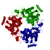


 PDBj
PDBj


 Assembly
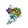


 Mass: 175.820 Da / Num. of mol.: 1 / Source method: obtained synthetically / Formula: Fe2S2
 Mass: 58.693 Da / Num. of mol.: 2 / Source method: obtained synthetically / Formula: Ni
 Mass: 35.453 Da / Num. of mol.: 1 / Source method: obtained synthetically / Formula: Cl
 Mass: 122.143 Da / Num. of mol.: 2 / Source method: obtained synthetically / Formula: C4H12NO3 / Comment: pH buffer*YM
 Mass: 92.094 Da / Num. of mol.: 2 / Source method: obtained synthetically / Formula: C3H8O3
 Sample preparation
 / Beamline: BL11-1 / Wavelength: 0.91837,0.97949,0.97895
 Processing