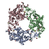
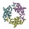
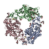
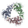
- PDB-1ww9: Crystal structure of the terminal oxygenase component of carbazol... -
+
Open data
ID or keywords:
Loading...
-
Basic information
Entry
Database: PDB / ID: 1ww9
Title
Crystal structure of the terminal oxygenase component of carbazole 1,9a-dioxygenase, a non-heme iron oxygenase system catalyzing the novel angular dioxygenation for carbazole and dioxin
Components
terminal oxygenase component of carbazole
Keywords
OXIDOREDUCTASE / Terminal oxygenase / Carbazole 1 / 9a-dioxygenase / Angular dioxygenase / Rieske non-heme iron oxygenase system
Function / homology
Function and homology information
: / organonitrogen compound catabolic process / 2 iron, 2 sulfur cluster binding / oxidoreductase activity / metal ion binding Similarity search - Function
N-terminal domain of TfIIb - #680 / Homotrimeric ring hydroxylase, catalytic domain / Homotrimeric ring hydroxylase / Naphthalene 1,2-dioxygenase Alpha Subunit; Chain A, domain 1 / Naphthalene 1,2-dioxygenase Alpha Subunit; Chain A, domain 1 / N-terminal domain of TfIIb - #10 / N-terminal domain of TfIIb / Rieske [2Fe-2S] iron-sulphur domain / Rieske [2Fe-2S] domain / Rieske [2Fe-2S] iron-sulfur domain profile. ...N-terminal domain of TfIIb - #680 / Homotrimeric ring hydroxylase, catalytic domain / Homotrimeric ring hydroxylase / Naphthalene 1,2-dioxygenase Alpha Subunit; Chain A, domain 1 / Naphthalene 1,2-dioxygenase Alpha Subunit; Chain A, domain 1 / N-terminal domain of TfIIb - #10 / N-terminal domain of TfIIb / Rieske [2Fe-2S] iron-sulphur domain / Rieske [2Fe-2S] domain / Rieske [2Fe-2S] iron-sulfur domain profile. / Rieske [2Fe-2S] iron-sulphur domain superfamily / Single Sheet / Alpha-Beta Complex / Mainly Beta / Alpha Beta Similarity search - Domain/homology
terminaloxygenasecomponentofcarbazole / Terminal oxygenase component of carbazole 1 / 9a-dioxygenase
Mass: 44910.738 Da / Num. of mol.: 1 Source method: isolated from a genetically manipulated source Source: (gene. exp.) Janthinobacterium sp. J3 (bacteria) / Plasmid: pEJ3AaC / Species (production host): Escherichia coli / Production host: Escherichia coli BL21(DE3) (bacteria) / Strain (production host): BL21(DE3) References: UniProt: Q84II6, Oxidoreductases; Acting on paired donors, with incorporation or reduction of molecular oxygen; With NADH or NADPH as one donor, and incorporation of two atoms of oxygen into the other donor
In the structure databanks used in Yorodumi, some data are registered as the other names, "COVID-19 virus" and "2019-nCoV". Here are the details of the virus and the list of structure data.
Jan 31, 2019. EMDB accession codes are about to change! (news from PDBe EMDB page)
EMDB accession codes are about to change! (news from PDBe EMDB page)
The allocation of 4 digits for EMDB accession codes will soon come to an end. Whilst these codes will remain in use, new EMDB accession codes will include an additional digit and will expand incrementally as the available range of codes is exhausted. The current 4-digit format prefixed with “EMD-” (i.e. EMD-XXXX) will advance to a 5-digit format (i.e. EMD-XXXXX), and so on. It is currently estimated that the 4-digit codes will be depleted around Spring 2019, at which point the 5-digit format will come into force.
The EM Navigator/Yorodumi systems omit the EMD- prefix.
Related info.:Q: What is EMD? / ID/Accession-code notation in Yorodumi/EM Navigator
Yorodumi is a browser for structure data from EMDB, PDB, SASBDB, etc.
This page is also the successor to EM Navigator detail page, and also detail information page/front-end page for Omokage search.
The word "yorodu" (or yorozu) is an old Japanese word meaning "ten thousand". "mi" (miru) is to see.
Related info.:EMDB / PDB / SASBDB / Comparison of 3 databanks / Yorodumi Search / Aug 31, 2016. New EM Navigator & Yorodumi / Yorodumi Papers / Jmol/JSmol / Function and homology information / Changes in new EM Navigator and Yorodumi
 Movie
Movie Controller

 Yorodumi
 Open data
 Basic information
 Components
 Keywords
 OXIDOREDUCTASE / Terminal oxygenase /
  Function and homology information
 Janthinobacterium sp. J3 (bacteria)
 Authors
 Citation
 Structure visualization
 Downloads & links
 Other downloads
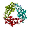


 PDBj
PDBj


 Assembly


 Mass: 55.845 Da / Num. of mol.: 1 / Source method: obtained synthetically / Formula: Fe
 Mass: 175.820 Da / Num. of mol.: 1 / Source method: obtained synthetically / Formula: Fe2S2
 Mass: 18.015 Da / Num. of mol.: 320 / Source method: isolated from a natural source / Formula: H2O
 Sample preparation
 / Beamline: BL41XU / Wavelength: 0.9794, 0.9796, 0.9820
 Processing