 Movie
Movie Controller


+ Open data

- Basic information
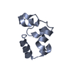
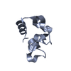
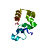
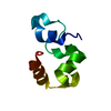
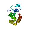
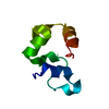
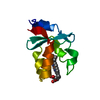
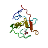
| Entry | Database: PDB / ID: 3myc | ||||||
|---|---|---|---|---|---|---|---|
| Title | Crystal Structure of HP67 H41F - P212121 | ||||||
 Components Components | Villin-1 | ||||||
 Keywords Keywords | STRUCTURAL PROTEIN / Protein / Helix / Hydrogen Bond | ||||||
| Function / homology |  Function and homology information Function and homology informationregulation of actin nucleation / cytoplasmic actin-based contraction involved in cell motility / lysophosphatidic acid binding / regulation of lamellipodium morphogenesis / filopodium tip / positive regulation of actin filament bundle assembly / actin filament severing / regulation of wound healing / actin filament capping / barbed-end actin filament capping ...regulation of actin nucleation / cytoplasmic actin-based contraction involved in cell motility / lysophosphatidic acid binding / regulation of lamellipodium morphogenesis / filopodium tip / positive regulation of actin filament bundle assembly / actin filament severing / regulation of wound healing / actin filament capping / barbed-end actin filament capping / actin polymerization or depolymerization / actin filament depolymerization / cellular response to hepatocyte growth factor stimulus / positive regulation of epithelial cell migration / actin filament bundle / microvillus / cysteine-type endopeptidase inhibitor activity involved in apoptotic process / cellular response to epidermal growth factor stimulus / ruffle / phosphatidylinositol-4,5-bisphosphate binding / actin filament polymerization / filopodium / response to bacterium / epidermal growth factor receptor signaling pathway / actin filament binding / actin cytoskeleton / lamellipodium / regulation of cell shape / positive regulation of cell migration / calcium ion binding / protein homodimerization activity / cytoplasmSimilarity search - Function | ||||||
| Biological species |  Gallus gallus (chicken) Gallus gallus (chicken) | ||||||
| Method | X-RAY DIFFRACTION / MOLECULAR REPLACEMENT / Resolution: 1.7 Å | ||||||
 Authors Authors | Brown, J.W. / Farelli, J.D. / McKnight, C.J. | ||||||
 Citation Citation | Journal: J.Mol.Biol. / Year: 2011 Title: On unsatisfied hydrogen bonds in the N-terminal subdomain of villin headpiece. Authors: Brown, J.W. / Farelli, J.D. / McKnight, C.J. | ||||||
| History |
|
- Structure visualization
| Structure viewer | Molecule: MolmilJmol/JSmol |
|---|
- Downloads & links
-Download
| PDBx/mmCIF format | 3myc.cif.gz | 26.9 KB | Display | PDBx/mmCIF format |
|---|---|---|---|---|
| PDB format | pdb3myc.ent.gz | 17.1 KB | Display | PDB format |
| PDBx/mmJSON format | 3myc.json.gz | Tree view | PDBx/mmJSON format | |
| Others |  Other downloads Other downloads |
-Validation report
| Arichive directory | https://data.pdbj.org/pub/pdb/validation_reports/my/3mycftp://data.pdbj.org/pub/pdb/validation_reports/my/3myc | HTTPS FTP |
|---|
-Related structure data


-Links
 PDBj
PDBj
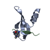
- Assembly
| Deposited unit | 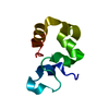
| ||||||||
|---|---|---|---|---|---|---|---|---|---|
| 1 |
| ||||||||
| Unit cell |
|
-Components
| #1: Protein | Mass: 7620.689 Da / Num. of mol.: 1 / Fragment: Villin Headpiece (UNP residues 760-826) / Mutation: H41F Source method: isolated from a genetically manipulated source Source: (gene. exp.) Gallus gallus (chicken) / Gene: VIL, VIL1 / Plasmid: pET-24A / Production host:  Escherichia coli (E. coli) / Strain (production host): BL21(DE3) / References: UniProt: P02640 Escherichia coli (E. coli) / Strain (production host): BL21(DE3) / References: UniProt: P02640 |
|---|---|
| #2: Water | ChemComp-HOH / Water Mass: 18.015 Da / Num. of mol.: 139 / Source method: isolated from a natural source / Formula: H2O Mass: 18.015 Da / Num. of mol.: 139 / Source method: isolated from a natural source / Formula: H2O |
-Experimental details
-Experiment
| Experiment | Method: X-RAY DIFFRACTION / Number of used crystals: 1 |
|---|
- Sample preparation
| Crystal | Density Matthews: 2.19 Å3/Da / Density % sol: 43.8 % |
|---|---|
| Crystal grow | Temperature: 293 K / Method: vapor diffusion, hanging drop / pH: 6.5 Details: 0.2 M Ammonium Sulfate, 0.1 M 2-Morphinoethanesulfonic acid, 30% Polyethyleneglycol 8,000, pH 6.5, VAPOR DIFFUSION, HANGING DROP, temperature 293K |
-Data collection
| Diffraction | Mean temperature: 200 K |
|---|---|
| Diffraction source | Source: ROTATING ANODE / Type: RIGAKU RU300 / Wavelength: 1.5418 Å |
| Detector | Type: RIGAKU RAXIS IV / Detector: IMAGE PLATE / Date: Mar 19, 2010 |
| Radiation | Protocol: SINGLE WAVELENGTH / Monochromatic (M) / Laue (L): M / Scattering type: x-ray |
| Radiation wavelength | Wavelength: 1.5418 Å / Relative weight: 1 |
| Reflection | Resolution: 1.7→13.9823 Å / Num. obs: 6978 / % possible obs: 93.85 % / Observed criterion σ(F): 0 / Observed criterion σ(I): 0 |
| Reflection shell | Resolution: 1.7→1.8311 Å / % possible all: 91 |
- Processing
| Software |
| ||||||||||||||||||||||||||||||||||||||||||
|---|---|---|---|---|---|---|---|---|---|---|---|---|---|---|---|---|---|---|---|---|---|---|---|---|---|---|---|---|---|---|---|---|---|---|---|---|---|---|---|---|---|---|---|
| Refinement | Method to determine structure: MOLECULAR REPLACEMENT / Resolution: 1.7→13.982 Å / SU ML: 0.11 / σ(F): 0.16 / Stereochemistry target values: ML
| ||||||||||||||||||||||||||||||||||||||||||
| Solvent computation | Shrinkage radii: 0.9 Å / VDW probe radii: 1.11 Å / Solvent model: FLAT BULK SOLVENT MODEL / Bsol: 42.827 Å2 / ksol: 0.355 e/Å3 | ||||||||||||||||||||||||||||||||||||||||||
| Displacement parameters |
| ||||||||||||||||||||||||||||||||||||||||||
| Refinement step | Cycle: LAST / Resolution: 1.7→13.982 Å
| ||||||||||||||||||||||||||||||||||||||||||
| Refine LS restraints |
| ||||||||||||||||||||||||||||||||||||||||||
| LS refinement shell |
|
