 Movie
Movie Controller

[English] 日本語
 Yorodumi
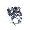
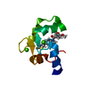
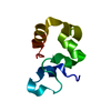
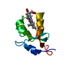
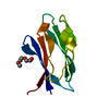
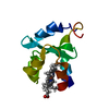
- PDB-1cxy: STRUCTURE AND CHARACTERIZATION OF ECTOTHIORHODOSPIRA VACUOLATA CY... -
+ Open data

- Basic information
| Entry | Database: PDB / ID: 1cxy | ||||||
|---|---|---|---|---|---|---|---|
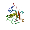
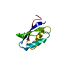
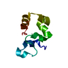
| Title | STRUCTURE AND CHARACTERIZATION OF ECTOTHIORHODOSPIRA VACUOLATA CYTOCHROME B558, A PROKARYOTIC HOMOLOGUE OF CYTOCHROME B5 | ||||||
 Components Components | CYTOCHROME B5 | ||||||
 Keywords Keywords | ELECTRON TRANSPORT / HELIX / BETA-STRAND | ||||||
| Function / homology |  Function and homology information Function and homology information | ||||||
| Biological species |  Ectothiorhodospira shaposhnikovii (bacteria) Ectothiorhodospira shaposhnikovii (bacteria) | ||||||
| Method | X-RAY DIFFRACTION / SYNCHROTRON / Resolution: 1.65 Å | ||||||
 Authors Authors | Kostanjevecki, V. / Leys, D. / Van Driessche, G. / Meyer, T.E. / Cusanovich, M.A. / Fischer, U. / Guisez, Y. / Van Beeumen, J. | ||||||
 Citation Citation | Journal: J.Biol.Chem. / Year: 1999 Title: Structure and characterization of Ectothiorhodospira vacuolata cytochrome b(558), a prokaryotic homologue of cytochrome b(5). Authors: Kostanjevecki, V. / Leys, D. / Van Driessche, G. / Meyer, T.E. / Cusanovich, M.A. / Fischer, U. / Guisez, Y. / Van Beeumen, J. | ||||||
| History |
|
- Structure visualization
| Structure viewer | Molecule: MolmilJmol/JSmol |
|---|
- Downloads & links
-Download
| PDBx/mmCIF format | 1cxy.cif.gz | 28.4 KB | Display | PDBx/mmCIF format |
|---|---|---|---|---|
| PDB format | pdb1cxy.ent.gz | 21.7 KB | Display | PDB format |
| PDBx/mmJSON format | 1cxy.json.gz | Tree view | PDBx/mmJSON format | |
| Others |  Other downloads Other downloads |
-Validation report
| Arichive directory | https://data.pdbj.org/pub/pdb/validation_reports/cx/1cxyftp://data.pdbj.org/pub/pdb/validation_reports/cx/1cxy | HTTPS FTP |
|---|
-Related structure data
| Similar structure data |
|---|
-Links
 PDBj
PDBj
- Assembly
| Deposited unit | 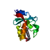
| ||||||||
|---|---|---|---|---|---|---|---|---|---|
| 1 |
| ||||||||
| Unit cell |
|
-Components
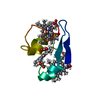
| #1: Protein | Mass: 10106.286 Da / Num. of mol.: 1 Source method: isolated from a genetically manipulated source Source: (gene. exp.) Ectothiorhodospira shaposhnikovii (bacteria)Production host: Escherichia coli (E. coli) / References: UniProt: P82291 |
|---|---|
| #2: Chemical | ChemComp-HEM / Heme B  Mass: 616.487 Da / Num. of mol.: 1 / Source method: obtained synthetically / Formula: C34H32FeN4O4 Mass: 616.487 Da / Num. of mol.: 1 / Source method: obtained synthetically / Formula: C34H32FeN4O4 |
| #3: Water | ChemComp-HOH / Water Mass: 18.015 Da / Num. of mol.: 166 / Source method: isolated from a natural source / Formula: H2O Mass: 18.015 Da / Num. of mol.: 166 / Source method: isolated from a natural source / Formula: H2O |
-Experimental details
-Experiment
| Experiment | Method: X-RAY DIFFRACTION / Number of used crystals: 1 |
|---|
- Sample preparation
| Crystal | Density Matthews: 2.82 Å3/Da / Density % sol: 56.34 % | ||||||||||||||||||||||||||||||
|---|---|---|---|---|---|---|---|---|---|---|---|---|---|---|---|---|---|---|---|---|---|---|---|---|---|---|---|---|---|---|---|
| Crystal grow | Temperature: 298 K / Method: vapor diffusion, hanging drop / pH: 5.6 Details: MES, NACL, pH 5.6, VAPOR DIFFUSION, HANGING DROP, temperature 298K | ||||||||||||||||||||||||||||||
| Crystal | *PLUS Density % sol: 63.8 % | ||||||||||||||||||||||||||||||
| Crystal grow | *PLUS pH: 7.4 | ||||||||||||||||||||||||||||||
| Components of the solutions | *PLUS
|
-Data collection
| Diffraction | Mean temperature: 100 K |
|---|---|
| Diffraction source | Source: SYNCHROTRON / Site: LURE  / Beamline: DW32 / Wavelength: 0.97 / Beamline: DW32 / Wavelength: 0.97 |
| Detector | Type: MARRESEARCH / Detector: IMAGE PLATE / Date: May 15, 1999 |
| Radiation | Protocol: SINGLE WAVELENGTH / Monochromatic (M) / Laue (L): M / Scattering type: x-ray |
| Radiation wavelength | Wavelength: 0.97 Å / Relative weight: 1 |
| Reflection | Resolution: 1.65→15 Å / Num. all: 92730 / Num. obs: 14370 / % possible obs: 99.6 % / Observed criterion σ(F): 1 / Observed criterion σ(I): 1 / Redundancy: 6.45 % / Biso Wilson estimate: 23.13 Å2 / Rmerge(I) obs: 0.045 / Net I/σ(I): 19.9 |
| Reflection shell | Resolution: 1.6→1.66 Å / Redundancy: 2.4 % / Rmerge(I) obs: 0.318 / % possible all: 99.7 |
| Reflection | *PLUS Num. measured all: 92730 / Rmerge(I) obs: 0.059 |
| Reflection shell | *PLUS % possible obs: 99.7 % |
- Processing
| Software |
| |||||||||||||||||||||||||
|---|---|---|---|---|---|---|---|---|---|---|---|---|---|---|---|---|---|---|---|---|---|---|---|---|---|---|
| Refinement | Resolution: 1.65→12 Å / σ(F): 0 / σ(I): 0 / Stereochemistry target values: ENGH & HUBER / Details: USE MAXIMUM LIKELIHOOD PROCEDURE
| |||||||||||||||||||||||||
| Refinement step | Cycle: LAST / Resolution: 1.65→12 Å
| |||||||||||||||||||||||||
| Software | *PLUS Name: REFMAC / Classification: refinement | |||||||||||||||||||||||||
| Refinement | *PLUS Rfactor all: 0.185 | |||||||||||||||||||||||||
| Solvent computation | *PLUS | |||||||||||||||||||||||||
| Displacement parameters | *PLUS | |||||||||||||||||||||||||
| Refine LS restraints | *PLUS
|
