 Movie
Movie Controller

[English] 日本語
 Yorodumi
- PDB-3mtk: X-Ray Structure of Diguanylate cyclase/phosphodiesterase from Cal... -
+ Open data

- Basic information
| Entry | Database: PDB / ID: 3mtk | ||||||
|---|---|---|---|---|---|---|---|
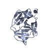
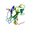
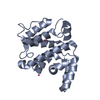
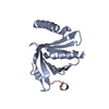
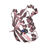
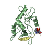
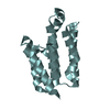
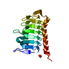
| Title | X-Ray Structure of Diguanylate cyclase/phosphodiesterase from Caldicellulosiruptor saccharolyticus, Northeast Structural Genomics Consortium Target ClR27C | ||||||
 Components Components | Diguanylate cyclase/phosphodiesterase | ||||||
 Keywords Keywords |  TRANSFERASE / Structural Genomics / PSI-2 / Protein Structure Initiative / Northeast Structural Genomics Consortium / NESG TRANSFERASE / Structural Genomics / PSI-2 / Protein Structure Initiative / Northeast Structural Genomics Consortium / NESG | ||||||
| Function / homology |  Function and homology information Function and homology informationPutative diguanylate phosphodiesterase / EAL domain / EAL domain superfamily / EAL domain / EAL domain profile. / Diguanylate cyclase, GGDEF domain / diguanylate cyclase / GGDEF domain profile. / GGDEF domain / Nucleotide cyclase ...Putative diguanylate phosphodiesterase / EAL domain / EAL domain superfamily / EAL domain / EAL domain profile. / Diguanylate cyclase, GGDEF domain / diguanylate cyclase / GGDEF domain profile. / GGDEF domain / Nucleotide cyclase / Reverse transcriptase/Diguanylate cyclase domain / PAS repeat profile. / PAS domain / PAS domain superfamily / Reverse transcriptase/Diguanylate cyclase domain / Alpha-Beta Plaits / 2-Layer Sandwich / Alpha BetaSimilarity search - Domain/homology | ||||||
| Biological species |   Caldicellulosiruptor saccharolyticus (bacteria) Caldicellulosiruptor saccharolyticus (bacteria) | ||||||
| Method | X-RAY DIFFRACTION / SYNCHROTRON / MAD / Resolution: 2.24 Å | ||||||
 Authors Authors | Kuzin, A. / Abashidze, M. / Seetharaman, J. / Sahdev, S. / Xiao, R. / Ciccosanti, C. / Wang, D. / Everett, J.K. / Nair, R. / Acton, T.B. ...Kuzin, A. / Abashidze, M. / Seetharaman, J. / Sahdev, S. / Xiao, R. / Ciccosanti, C. / Wang, D. / Everett, J.K. / Nair, R. / Acton, T.B. / Rost, B. / Montelione, G.T. / Tong, L. / Hunt, J.F. / Northeast Structural Genomics Consortium (NESG) | ||||||
 Citation Citation | Journal: To be Published Title: Northeast Structural Genomics Consortium Target ClR27C Authors: Kuzin, A. / Abashidze, M. / Seetharaman, J. / Sahdev, S. / Xiao, R. / Ciccosanti, C. / Wang, D. / Everett, J.K. / Nair, R. / Acton, T.B. / Rost, B. / Montelione, G.T. / Tong, L. / Hunt, J.F. | ||||||
| History |
|
- Structure visualization
| Structure viewer | Molecule: MolmilJmol/JSmol |
|---|
- Downloads & links
-Download
| PDBx/mmCIF format | 3mtk.cif.gz | 73.6 KB | Display | PDBx/mmCIF format |
|---|---|---|---|---|
| PDB format | pdb3mtk.ent.gz | 58.9 KB | Display | PDB format |
| PDBx/mmJSON format | 3mtk.json.gz | Tree view | PDBx/mmJSON format | |
| Others |  Other downloads Other downloads |
-Validation report
| Arichive directory | https://data.pdbj.org/pub/pdb/validation_reports/mt/3mtkftp://data.pdbj.org/pub/pdb/validation_reports/mt/3mtk | HTTPS FTP |
|---|
-Related structure data
| Similar structure data | |
|---|---|
| Other databases |


-Links
 PDBj
PDBj


- Assembly
| Deposited unit | 
| ||||||||
|---|---|---|---|---|---|---|---|---|---|
| 1 | 
| ||||||||
| 2 | 
| ||||||||
| 3 |
| ||||||||
| Unit cell |
| ||||||||
| Details | monomer,24.24 kD,97.6% |
-Components
| #1: Protein | Mass: 20715.357 Da / Num. of mol.: 2 Source method: isolated from a genetically manipulated source Source: (gene. exp.) Caldicellulosiruptor saccharolyticus (bacteria)Strain: ATCC 43494 / DSM 8903 / Gene: Csac_0821 / Plasmid: pET 21-23C / Production host: Escherichia coli (E. coli) / Strain (production host): BL21(DE3)+ Magic / References: UniProt: A4XHQ4#2: Water | ChemComp-HOH / | Water Mass: 18.015 Da / Num. of mol.: 38 / Source method: isolated from a natural source / Formula: H2O Mass: 18.015 Da / Num. of mol.: 38 / Source method: isolated from a natural source / Formula: H2O |
|---|
-Experimental details
-Experiment
| Experiment | Method: X-RAY DIFFRACTION / Number of used crystals: 1 |
|---|
- Sample preparation
| Crystal | Density Matthews: 2.12 Å3/Da / Density % sol: 42.03 % |
|---|---|
| Crystal grow | pH: 7.5 Details: Protein solution: 100mM NaCl, 5mM DTT, 0.02% NaN3, 10mM Tris-HCl pH 7.5. Reservoir solution:, VAPOR DIFFUSION, HANGING DROP |
-Data collection
| Diffraction | Mean temperature: 100 K | ||||||||||||
|---|---|---|---|---|---|---|---|---|---|---|---|---|---|
| Diffraction source | Source: SYNCHROTRON / Site: NSLS  / Beamline: X4A / Wavelength: 0.97910, 0.97924, 0.96791 / Beamline: X4A / Wavelength: 0.97910, 0.97924, 0.96791 | ||||||||||||
| Detector | Type: ADSC QUANTUM 4 / Detector: CCD / Date: Apr 9, 2010 / Details: MIRRORS | ||||||||||||
| Radiation | Monochromator: SI 111 CHANNEL / Protocol: MAD / Monochromatic (M) / Laue (L): M / Scattering type: x-ray | ||||||||||||
| Radiation wavelength |
| ||||||||||||
| Reflection | Resolution: 2.24→50 Å / Num. obs: 32644 / % possible obs: 99.5 % / Observed criterion σ(I): -3 / Redundancy: 3.7 % / Rmerge(I) obs: 0.058 / Net I/σ(I): 32.3 | ||||||||||||
| Reflection shell | Resolution: 2.24→2.32 Å / Redundancy: 3.2 % / Rmerge(I) obs: 0.337 / Mean I/σ(I) obs: 7.3 / % possible all: 96.8 |
- Processing
| Software |
| ||||||||||||||||||||||||||||||||||||||||||||||||||||||||||||
|---|---|---|---|---|---|---|---|---|---|---|---|---|---|---|---|---|---|---|---|---|---|---|---|---|---|---|---|---|---|---|---|---|---|---|---|---|---|---|---|---|---|---|---|---|---|---|---|---|---|---|---|---|---|---|---|---|---|---|---|---|---|
| Refinement | Method to determine structure: MAD / Resolution: 2.24→20 Å / σ(F): 0 / Stereochemistry target values: ENGH & HUBER
| ||||||||||||||||||||||||||||||||||||||||||||||||||||||||||||
| Solvent computation | Bsol: 63.57 Å2 | ||||||||||||||||||||||||||||||||||||||||||||||||||||||||||||
| Displacement parameters | Biso mean: 51.72 Å2
| ||||||||||||||||||||||||||||||||||||||||||||||||||||||||||||
| Refinement step | Cycle: LAST / Resolution: 2.24→20 Å
| ||||||||||||||||||||||||||||||||||||||||||||||||||||||||||||
| Refine LS restraints |
| ||||||||||||||||||||||||||||||||||||||||||||||||||||||||||||
| Xplor file |
|
