 Movie
Movie Controller


+ Open data

- Basic information
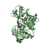
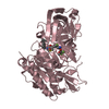
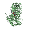
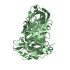
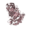
| Entry | Database: PDB / ID: 3msl | ||||||
|---|---|---|---|---|---|---|---|
| Title | Fragment Based Discovery and Optimisation of BACE-1 Inhibitors | ||||||
 Components Components | Beta-secretase 1 | ||||||
 Keywords Keywords | HYDROLASE/HYDROLASE INHIBITOR / PROTEASE / ALZHEIMER'S DISEASE / ASPARTIC PROTEASE / ASPARTYL PROTEASE / BASE / BETA-SECRETASE / GLYCOPROTEIN / HYDROLASE / MEMAPSIN 2 / AMYLOID PRECURSOR PROTEIN SECRETASES / ASPARTIC ENDOPEPTIDASES / FRAGMENT-BASED DRUG DESIGN / FLUORESCENCE POLARISATION / TRANSMEMBRANE / ZYMOGEN / HYDROLASE-HYDROLASE INHIBITOR complex | ||||||
| Function / homology |  Function and homology informationmemapsin 2 / Golgi-associated vesicle lumen / signaling receptor ligand precursor processing / beta-aspartyl-peptidase activity / amyloid precursor protein catabolic process / amyloid-beta formation / membrane protein ectodomain proteolysis / cellular response to manganese ion / amyloid-beta metabolic process / prepulse inhibition ...memapsin 2 / Golgi-associated vesicle lumen / signaling receptor ligand precursor processing / beta-aspartyl-peptidase activity / amyloid precursor protein catabolic process / amyloid-beta formation / membrane protein ectodomain proteolysis / cellular response to manganese ion / amyloid-beta metabolic process / prepulse inhibition / detection of mechanical stimulus involved in sensory perception of pain / cellular response to copper ion / presynaptic modulation of chemical synaptic transmission / hippocampal mossy fiber to CA3 synapse / multivesicular body / response to lead ion / trans-Golgi network / recycling endosome / protein processing / cellular response to amyloid-beta / positive regulation of neuron apoptotic process / synaptic vesicle / late endosome / peptidase activity / amyloid-beta binding / endopeptidase activity / amyloid fibril formation / lysosome / aspartic-type endopeptidase activity / early endosome / endosome membrane / endosome / membrane raft / Amyloid fiber formation / endoplasmic reticulum lumen / axon / neuronal cell body / dendrite / Golgi apparatus / enzyme binding / cell surface / proteolysis / membrane / plasma membrane Function and homology informationmemapsin 2 / Golgi-associated vesicle lumen / signaling receptor ligand precursor processing / beta-aspartyl-peptidase activity / amyloid precursor protein catabolic process / amyloid-beta formation / membrane protein ectodomain proteolysis / cellular response to manganese ion / amyloid-beta metabolic process / prepulse inhibition ...memapsin 2 / Golgi-associated vesicle lumen / signaling receptor ligand precursor processing / beta-aspartyl-peptidase activity / amyloid precursor protein catabolic process / amyloid-beta formation / membrane protein ectodomain proteolysis / cellular response to manganese ion / amyloid-beta metabolic process / prepulse inhibition / detection of mechanical stimulus involved in sensory perception of pain / cellular response to copper ion / presynaptic modulation of chemical synaptic transmission / hippocampal mossy fiber to CA3 synapse / multivesicular body / response to lead ion / trans-Golgi network / recycling endosome / protein processing / cellular response to amyloid-beta / positive regulation of neuron apoptotic process / synaptic vesicle / late endosome / peptidase activity / amyloid-beta binding / endopeptidase activity / amyloid fibril formation / lysosome / aspartic-type endopeptidase activity / early endosome / endosome membrane / endosome / membrane raft / Amyloid fiber formation / endoplasmic reticulum lumen / axon / neuronal cell body / dendrite / Golgi apparatus / enzyme binding / cell surface / proteolysis / membrane / plasma membraneSimilarity search - Function | ||||||
| Biological species |  Homo sapiens (human) Homo sapiens (human) | ||||||
| Method | X-RAY DIFFRACTION / SYNCHROTRON / MOLECULAR REPLACEMENT / Resolution: 2.4 Å | ||||||
 Authors Authors | Smith, M. / Madden, J. / Barker, J. | ||||||
 Citation Citation | Journal: Bioorg.Med.Chem.Lett. / Year: 2010 Title: Fragment-based discovery and optimization of BACE1 inhibitors. Authors: Madden, J. / Dod, J.R. / Godemann, R. / Kraemer, J. / Smith, M. / Biniszkiewicz, M. / Hallett, D.J. / Barker, J. / Dyekjaer, J.D. / Hesterkamp, T. | ||||||
| History |
|
- Structure visualization
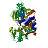
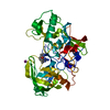
| Structure viewer | Molecule: MolmilJmol/JSmol |
|---|
- Downloads & links
-Download
| PDBx/mmCIF format | 3msl.cif.gz | 169.8 KB | Display | PDBx/mmCIF format |
|---|---|---|---|---|
| PDB format | pdb3msl.ent.gz | 133.4 KB | Display | PDB format |
| PDBx/mmJSON format | 3msl.json.gz | Tree view | PDBx/mmJSON format | |
| Others |  Other downloads Other downloads |
-Validation report
| Arichive directory | https://data.pdbj.org/pub/pdb/validation_reports/ms/3mslftp://data.pdbj.org/pub/pdb/validation_reports/ms/3msl | HTTPS FTP |
|---|
-Related structure data
| Related structure data |  3msjC  3mskC  3s2oC 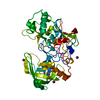 1w50S C: citing same article ( S: Starting model for refinement |
|---|---|
| Similar structure data |


-Links
 PDBj
PDBj

- Assembly
| Deposited unit | 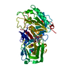
| ||||||||
|---|---|---|---|---|---|---|---|---|---|
| 1 |
| ||||||||
| Unit cell |
|
-Components
| #1: Protein | / Beta-site amyloid precursor protein cleaving enzyme 1 / Beta-site APP cleaving enzyme 1 / Membrane- ...Beta-site amyloid precursor protein cleaving enzyme 1 / Beta-site APP cleaving enzyme 1 / Membrane-associated aspartic protease 2 / Memapsin-2 / Aspartyl protease 2 / Asp 2 / ASP2 Mass: 45434.957 Da / Num. of mol.: 1 / Fragment: UNP RESIDUES 48-453 Source method: isolated from a genetically manipulated source Source: (gene. exp.) Homo sapiens (human) / Gene: BACE, BACE1, KIAA1149 / Plasmid: PET11A / Production host:  Escherichia coli (E. coli) / Strain (production host): BL21 (DE3) / References: UniProt: P56817, memapsin 2 Escherichia coli (E. coli) / Strain (production host): BL21 (DE3) / References: UniProt: P56817, memapsin 2 | ||||
|---|---|---|---|---|---|
| #2: Chemical | Iodide  Mass: 126.904 Da / Num. of mol.: 2 / Source method: obtained synthetically / Formula: I Mass: 126.904 Da / Num. of mol.: 2 / Source method: obtained synthetically / Formula: I#3: Chemical | ChemComp-EV5 / ( |   Mass: 362.897 Da / Num. of mol.: 1 / Source method: obtained synthetically / Formula: C19H27ClN4O Mass: 362.897 Da / Num. of mol.: 1 / Source method: obtained synthetically / Formula: C19H27ClN4O#4: Water | ChemComp-HOH / | Water Mass: 18.015 Da / Num. of mol.: 159 / Source method: isolated from a natural source / Formula: H2O Mass: 18.015 Da / Num. of mol.: 159 / Source method: isolated from a natural source / Formula: H2O |
-Experimental details
-Experiment
| Experiment | Method: X-RAY DIFFRACTION / Number of used crystals: 1 |
|---|
- Sample preparation
| Crystal | Density Matthews: 2.76 Å3/Da / Density % sol: 55.37 % |
|---|---|
| Crystal grow | Temperature: 273 K / pH: 6.6 Details: 20% PEG 5000MME 180mM Ammonium Iodide and 180mM Sodium Citrate pH6.6 , VAPOR DIFFUSION, HANGING DROP, temperature 273K |
-Data collection
| Diffraction | Mean temperature: 100 K |
|---|---|
| Diffraction source | Source: SYNCHROTRON / Site: Diamond  / Beamline: I04 / Wavelength: 1.0332 / Beamline: I04 / Wavelength: 1.0332 |
| Detector | Type: ADSC QUANTUM 315r / Detector: CCD / Date: Sep 17, 2008 |
| Radiation | Monochromator: DOUBLE CRYSTAL / Protocol: SINGLE WAVELENGTH / Monochromatic (M) / Laue (L): M / Scattering type: x-ray |
| Radiation wavelength | Wavelength: 1.0332 Å / Relative weight: 1 |
| Reflection | Resolution: 2.4→61.2 Å / Num. obs: 17067 / % possible obs: 86.3 % / Observed criterion σ(I): 2 / Redundancy: 11.89 % / Biso Wilson estimate: 40.5 Å2 / Rmerge(I) obs: 0.102 / Rsym value: 0.106 / Net I/σ(I): 12.9 |
| Reflection shell | Resolution: 2.4→2.46 Å / Redundancy: 11.7 % / Rmerge(I) obs: 0.287 / Mean I/σ(I) obs: 4.3 / Rsym value: 0.131 / % possible all: 89.5 |
- Processing
| Software |
| ||||||||||||||||||||||||||||||||||||||||||||||||||||||||||||||||||||||||||||||||||||||||||||||||||||||||||||||||||||||||||||||||||||||||||||||||||||||||||||||||||||||||||
|---|---|---|---|---|---|---|---|---|---|---|---|---|---|---|---|---|---|---|---|---|---|---|---|---|---|---|---|---|---|---|---|---|---|---|---|---|---|---|---|---|---|---|---|---|---|---|---|---|---|---|---|---|---|---|---|---|---|---|---|---|---|---|---|---|---|---|---|---|---|---|---|---|---|---|---|---|---|---|---|---|---|---|---|---|---|---|---|---|---|---|---|---|---|---|---|---|---|---|---|---|---|---|---|---|---|---|---|---|---|---|---|---|---|---|---|---|---|---|---|---|---|---|---|---|---|---|---|---|---|---|---|---|---|---|---|---|---|---|---|---|---|---|---|---|---|---|---|---|---|---|---|---|---|---|---|---|---|---|---|---|---|---|---|---|---|---|---|---|---|---|---|
| Refinement | Method to determine structure: MOLECULAR REPLACEMENT Starting model: PDB ENTRY 1W50 Resolution: 2.4→50.1 Å / Cor.coef. Fo:Fc: 0.912 / Cor.coef. Fo:Fc free: 0.913 / SU B: 15.199 / SU ML: 0.165 / Cross valid method: THROUGHOUT / σ(F): 2 / ESU R Free: 0.258 / Stereochemistry target values: MAXIMUM LIKELIHOOD / Details: HYDROGENS HAVE BEEN ADDED IN THE RIDING POSITIONS
| ||||||||||||||||||||||||||||||||||||||||||||||||||||||||||||||||||||||||||||||||||||||||||||||||||||||||||||||||||||||||||||||||||||||||||||||||||||||||||||||||||||||||||
| Solvent computation | Ion probe radii: 0.8 Å / Shrinkage radii: 0.8 Å / VDW probe radii: 1.4 Å / Solvent model: MASK | ||||||||||||||||||||||||||||||||||||||||||||||||||||||||||||||||||||||||||||||||||||||||||||||||||||||||||||||||||||||||||||||||||||||||||||||||||||||||||||||||||||||||||
| Displacement parameters | Biso mean: 24.11 Å2
| ||||||||||||||||||||||||||||||||||||||||||||||||||||||||||||||||||||||||||||||||||||||||||||||||||||||||||||||||||||||||||||||||||||||||||||||||||||||||||||||||||||||||||
| Refinement step | Cycle: LAST / Resolution: 2.4→50.1 Å
| ||||||||||||||||||||||||||||||||||||||||||||||||||||||||||||||||||||||||||||||||||||||||||||||||||||||||||||||||||||||||||||||||||||||||||||||||||||||||||||||||||||||||||
| Refine LS restraints |
| ||||||||||||||||||||||||||||||||||||||||||||||||||||||||||||||||||||||||||||||||||||||||||||||||||||||||||||||||||||||||||||||||||||||||||||||||||||||||||||||||||||||||||
| LS refinement shell | Resolution: 2.4→2.46 Å / Total num. of bins used: 20
| ||||||||||||||||||||||||||||||||||||||||||||||||||||||||||||||||||||||||||||||||||||||||||||||||||||||||||||||||||||||||||||||||||||||||||||||||||||||||||||||||||||||||||
| Refinement TLS params. | Method: refined / Origin x: 14.6464 Å / Origin y: -40.8946 Å / Origin z: -0.6597 Å
|
