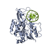
 Movie
Movie Controller


+ Open data

- Basic information
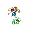
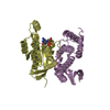
| Entry | Database: PDB / ID: 3mql | ||||||
|---|---|---|---|---|---|---|---|
| Title | Crystal structure of the fibronectin 6FnI1-2FnII7FnI fragment | ||||||
 Components Components | Fibronectin 1 | ||||||
 Keywords Keywords | CELL ADHESION / fibronectin / collagen / GBD / Fn2 / Fn1 | ||||||
| Function / homology |  Function and homology information Function and homology informationneutrophil degranulation / negative regulation of monocyte activation / calcium-independent cell-matrix adhesion / negative regulation of transforming growth factor beta production / Fibronectin matrix formation / Extracellular matrix organization / positive regulation of substrate-dependent cell migration, cell attachment to substrate / neural crest cell migration involved in autonomic nervous system development / peptidase activator activity / fibrinogen complex ...neutrophil degranulation / negative regulation of monocyte activation / calcium-independent cell-matrix adhesion / negative regulation of transforming growth factor beta production / Fibronectin matrix formation / Extracellular matrix organization / positive regulation of substrate-dependent cell migration, cell attachment to substrate / neural crest cell migration involved in autonomic nervous system development / peptidase activator activity / fibrinogen complex / peptide cross-linking / integrin activation / ALK mutants bind TKIs / poly(A) binding / cell-substrate junction assembly / U2-type catalytic step 2 spliceosome / biological process involved in interaction with symbiont / Molecules associated with elastic fibres / proteoglycan binding / extracellular matrix structural constituent / MET activates PTK2 signaling / cyclosporin A binding / Syndecan interactions / p130Cas linkage to MAPK signaling for integrins / endodermal cell differentiation / GRB2:SOS provides linkage to MAPK signaling for Integrins / Non-integrin membrane-ECM interactions / basement membrane / endoplasmic reticulum-Golgi intermediate compartment / ECM proteoglycans / positive regulation of axon extension / Integrin cell surface interactions / transcription-coupled nucleotide-excision repair / positive regulation of viral genome replication / protein peptidyl-prolyl isomerization / collagen binding / Degradation of the extracellular matrix / catalytic step 2 spliceosome / Integrin signaling / substrate adhesion-dependent cell spreading / mRNA Splicing - Major Pathway / cell-matrix adhesion / extracellular matrix / regulation of ERK1 and ERK2 cascade / platelet alpha granule lumen / peptidylprolyl isomerase / peptidyl-prolyl cis-trans isomerase activity / acute-phase response / integrin-mediated signaling pathway / Cell surface interactions at the vascular wall / Post-translational protein phosphorylation / regulation of protein phosphorylation / Signaling by high-kinase activity BRAF mutants / wound healing / MAP2K and MAPK activation / Signaling by ALK fusions and activated point mutants / Transcription-Coupled Nucleotide Excision Repair (TC-NER) / Formation of TC-NER Pre-Incision Complex / response to wounding / mRNA splicing, via spliceosome / Dual incision in TC-NER / Gap-filling DNA repair synthesis and ligation in TC-NER / GPER1 signaling / Signaling by RAF1 mutants / Signaling by moderate kinase activity BRAF mutants / Paradoxical activation of RAF signaling by kinase inactive BRAF / Signaling downstream of RAS mutants / Regulation of Insulin-like Growth Factor (IGF) transport and uptake by Insulin-like Growth Factor Binding Proteins (IGFBPs) / positive regulation of fibroblast proliferation / Signaling by BRAF and RAF1 fusions / integrin binding / protein folding / Platelet degranulation / heparin binding / nervous system development / heart development / regulation of cell shape / secretory granule lumen / Interleukin-4 and Interleukin-13 signaling / angiogenesis / collagen-containing extracellular matrix / protease binding / ficolin-1-rich granule lumen / blood microparticle / positive regulation of phosphatidylinositol 3-kinase/protein kinase B signal transduction / cell adhesion / nuclear speck / apical plasma membrane / endoplasmic reticulum lumen / signaling receptor binding / mRNA binding / Neutrophil degranulation / positive regulation of cell population proliferation / regulation of DNA-templated transcription / positive regulation of gene expression / extracellular space / RNA binding / extracellular exosome / extracellular region / nucleoplasmSimilarity search - Function | ||||||
| Biological species |  Homo sapiens (human) Homo sapiens (human) | ||||||
| Method | X-RAY DIFFRACTION / SYNCHROTRON / MOLECULAR REPLACEMENT / Resolution: 3.004 Å | ||||||
 Authors Authors | Erat, M.C. / Campbell, I.D. / Vakonakis, I. | ||||||
 Citation Citation | Journal: J.Biol.Chem. / Year: 2010 Title: Implications for collagen binding from the crystallographic structure of fibronectin 6FnI1-2FnII7FnI Authors: Erat, M.C. / Schwarz-Linek, U. / Pickford, A.R. / Farndale, R.W. / Campbell, I.D. / Vakonakis, I. | ||||||
| History |
|
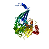
- Structure visualization
| Structure viewer | Molecule: MolmilJmol/JSmol |
|---|
- Downloads & links
-Download
| PDBx/mmCIF format | 3mql.cif.gz | 99.4 KB | Display | PDBx/mmCIF format |
|---|---|---|---|---|
| PDB format | pdb3mql.ent.gz | 75.5 KB | Display | PDB format |
| PDBx/mmJSON format | 3mql.json.gz | Tree view | PDBx/mmJSON format | |
| Others |  Other downloads Other downloads |
-Validation report
| Arichive directory | https://data.pdbj.org/pub/pdb/validation_reports/mq/3mqlftp://data.pdbj.org/pub/pdb/validation_reports/mq/3mql | HTTPS FTP |
|---|
-Related structure data
| Related structure data | 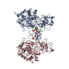 1eakS  1h8pS  1l6jS  2cg7S 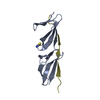 2rkyS 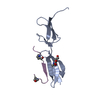 3ejhS S: Starting model for refinement |
|---|---|
| Similar structure data | |
| Other databases |
|


-Links
 PDBj
PDBj


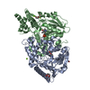
- Assembly
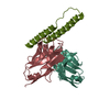
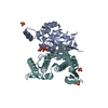
| Deposited unit | 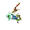
| ||||||||
|---|---|---|---|---|---|---|---|---|---|
| 1 |
| ||||||||
| Unit cell |
| ||||||||
| Components on special symmetry positions |
|
-Components
| #1: Protein | / Fibronectin 1 / isoform CRA_g Mass: 24874.469 Da / Num. of mol.: 1 / Fragment: 6FnI1-2FnII7FnI, UNP residues 305-515 / Mutation: H307D Source method: isolated from a genetically manipulated source Details: gene integrated in the AOX1 locus / Source: (gene. exp.) Homo sapiens (human) / Description: genomic integration / Gene: FN1, hCG_1813428 / Production host:  Pichia pastoris (fungus) / Strain (production host): X33 / References: UniProt: B7ZLF0, UniProt: P02751*PLUS Pichia pastoris (fungus) / Strain (production host): X33 / References: UniProt: B7ZLF0, UniProt: P02751*PLUS | ||
|---|---|---|---|
| #2: Sugar | ChemComp-NAG / N-Acetylglucosamine  Type: D-saccharide, beta linking / Mass: 221.208 Da / Num. of mol.: 1 Type: D-saccharide, beta linking / Mass: 221.208 Da / Num. of mol.: 1Source method: isolated from a genetically manipulated source Formula: C8H15NO6 | ||
| #3: Chemical | ChemComp-MPD / (2-Methyl-2,4-pentanediol  Mass: 118.174 Da / Num. of mol.: 1 / Source method: obtained synthetically / Formula: C6H14O2 / Comment: precipitant*YM Mass: 118.174 Da / Num. of mol.: 1 / Source method: obtained synthetically / Formula: C6H14O2 / Comment: precipitant*YM | ||
| #4: Chemical | 2-Methyl-2,4-pentanediol  Mass: 118.174 Da / Num. of mol.: 2 / Source method: obtained synthetically / Formula: C6H14O2 / Comment: precipitant*YM Mass: 118.174 Da / Num. of mol.: 2 / Source method: obtained synthetically / Formula: C6H14O2 / Comment: precipitant*YM#5: Water | ChemComp-HOH / | Water Mass: 18.015 Da / Num. of mol.: 2 / Source method: isolated from a natural source / Formula: H2O Mass: 18.015 Da / Num. of mol.: 2 / Source method: isolated from a natural source / Formula: H2O |
-Experimental details
-Experiment
| Experiment | Method: X-RAY DIFFRACTION / Number of used crystals: 1 |
|---|
- Sample preparation
| Crystal | Density Matthews: 2.77 Å3/Da / Density % sol: 55.62 % |
|---|---|
| Crystal grow | Temperature: 293 K / Method: vapor diffusion, sitting drop / pH: 7 Details: 1:1 drops of of protein in 100 mM NaCl, 10mM HEPES pH 7.2 buffer at 17.5 mg/ml concentration, and a 0.2 M (NH4)H2PO4, 0.1 M Tris-Cl pH 8.5, 50% MPD buffer, VAPOR DIFFUSION, SITTING DROP, temperature 293K |
-Data collection
| Diffraction | Mean temperature: 100 K |
|---|---|
| Diffraction source | Source: SYNCHROTRON / Site: SLS  / Beamline: X06DA / Wavelength: 1 Å / Beamline: X06DA / Wavelength: 1 Å |
| Detector | Type: MARMOSAIC 225 mm CCD / Detector: CCD / Date: Feb 5, 2010 |
| Radiation | Monochromator: Bartels monochromator with dual channel cut crystals Protocol: SINGLE WAVELENGTH / Monochromatic (M) / Laue (L): M / Scattering type: x-ray |
| Radiation wavelength | Wavelength: 1 Å / Relative weight: 1 |
| Reflection | Resolution: 3.004→51.974 Å / Num. obs: 5835 / % possible obs: 99.7 % / Observed criterion σ(F): 0 / Observed criterion σ(I): 0 / Redundancy: 7.1 % / Rsym value: 0.055 / Net I/σ(I): 27 |
| Reflection shell | Resolution: 3→3.17 Å / Redundancy: 7.1 % / Rmerge(I) obs: 0.473 / Mean I/σ(I) obs: 1.7 / Rsym value: 0.473 / % possible all: 98.6 |
- Processing
| Software |
| ||||||||||||||||||||||||||||||||||||||||
|---|---|---|---|---|---|---|---|---|---|---|---|---|---|---|---|---|---|---|---|---|---|---|---|---|---|---|---|---|---|---|---|---|---|---|---|---|---|---|---|---|---|
| Refinement | Method to determine structure: MOLECULAR REPLACEMENT Starting model: INDIVIDUAL DOMAINS FROM PDB ENTRIES 2CG7, 2RKY, 3EJH, 1EAK, 1H8P, 1L6J Resolution: 3.004→51.973 Å / Occupancy max: 1 / Occupancy min: 1 / SU ML: 0.35 / σ(F): 1.4 / Phase error: 23.3 / Stereochemistry target values: ML Details: 1. Used one TLS restraint for entire polypeptide chain; 2. SF file contains Friedel pairs.
| ||||||||||||||||||||||||||||||||||||||||
| Solvent computation | Shrinkage radii: 0.9 Å / VDW probe radii: 1.11 Å / Solvent model: FLAT BULK SOLVENT MODEL / Bsol: 52.177 Å2 / ksol: 0.3 e/Å3 | ||||||||||||||||||||||||||||||||||||||||
| Displacement parameters | Biso max: 287.1 Å2 / Biso min: 35.73 Å2
| ||||||||||||||||||||||||||||||||||||||||
| Refinement step | Cycle: LAST / Resolution: 3.004→51.973 Å
| ||||||||||||||||||||||||||||||||||||||||
| Refine LS restraints |
| ||||||||||||||||||||||||||||||||||||||||
| LS refinement shell |
| ||||||||||||||||||||||||||||||||||||||||
| Refinement TLS params. | Method: refined / Origin x: -2.5081 Å / Origin y: -23.3915 Å / Origin z: 36.3732 Å
| ||||||||||||||||||||||||||||||||||||||||
| Refinement TLS group | Selection details: chain A |
