 Movie
Movie Controller

[English] 日本語
 Yorodumi
- PDB-3mii: Crystal structure of Y0R391Cp/HSP33 from Saccharomyces cerevisiae -
+ Open data

- Basic information
| Entry | Database: PDB / ID: 3mii | ||||||
|---|---|---|---|---|---|---|---|
| Title | Crystal structure of Y0R391Cp/HSP33 from Saccharomyces cerevisiae | ||||||
 Components Components | Probable chaperone protein HSP33 | ||||||
 Keywords Keywords |  HYDROLASE / heat shock protein HYDROLASE / heat shock protein | ||||||
| Function / homology |  Function and homology informationD-lactate dehydratase / glyoxalase III activity / methylglyoxal catabolic process to D-lactate via S-lactoyl-glutathione / cellular response to nutrient levels / chaperone-mediated protein folding / protein folding chaperone / P-body / cytoplasm Function and homology informationD-lactate dehydratase / glyoxalase III activity / methylglyoxal catabolic process to D-lactate via S-lactoyl-glutathione / cellular response to nutrient levels / chaperone-mediated protein folding / protein folding chaperone / P-body / cytoplasmSimilarity search - Function | ||||||
| Biological species |  Saccharomyces cerevisiae (brewer's yeast) Saccharomyces cerevisiae (brewer's yeast) | ||||||
| Method | X-RAY DIFFRACTION / MOLECULAR REPLACEMENT / Resolution: 2.4 Å | ||||||
 Authors Authors | Guo, P.-C. / Zhou, Y.-Y. / Zhou, C.-Z. / Li, W.-F. | ||||||
 Citation Citation | Journal: Acta Crystallogr.,Sect.F / Year: 2010 Title: Structure of Hsp33/YOR391Cp from the yeast Saccharomyces cerevisiae Authors: Guo, P.-C. / Zhou, Y.-Y. / Ma, X.-X. / Li, W.-F. | ||||||
| History |
|
- Structure visualization
| Structure viewer | Molecule: MolmilJmol/JSmol |
|---|
- Downloads & links
-Download
| PDBx/mmCIF format | 3mii.cif.gz | 105.9 KB | Display | PDBx/mmCIF format |
|---|---|---|---|---|
| PDB format | pdb3mii.ent.gz | 80.9 KB | Display | PDB format |
| PDBx/mmJSON format | 3mii.json.gz | Tree view | PDBx/mmJSON format | |
| Others |  Other downloads Other downloads |
-Validation report
| Arichive directory | https://data.pdbj.org/pub/pdb/validation_reports/mi/3miiftp://data.pdbj.org/pub/pdb/validation_reports/mi/3mii | HTTPS FTP |
|---|
-Related structure data
| Related structure data |  1qvzS S: Starting model for refinement |
|---|---|
| Similar structure data |


-Links
 PDBj
PDBj

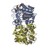
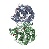
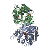
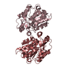
- Assembly
| Deposited unit | 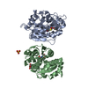
| ||||||||
|---|---|---|---|---|---|---|---|---|---|
| 1 |
| ||||||||
| 2 | 
| ||||||||
| 3 | 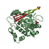
| ||||||||
| Unit cell |
|
-Components
| #1: Protein | Mass: 26992.549 Da / Num. of mol.: 2 Source method: isolated from a genetically manipulated source Source: (gene. exp.) Saccharomyces cerevisiae (brewer's yeast)Strain: S288C / Gene: YOR091C / Plasmid: pET28 / Production host:  Escherichia coli (E. coli) / Strain (production host): BL21(DE3) / References: UniProt: Q08914, Hydrolases; Glycosylases Escherichia coli (E. coli) / Strain (production host): BL21(DE3) / References: UniProt: Q08914, Hydrolases; Glycosylases#2: Chemical | Polyethylene glycol  Mass: 150.173 Da / Num. of mol.: 2 / Source method: obtained synthetically / Formula: C6H14O4 Mass: 150.173 Da / Num. of mol.: 2 / Source method: obtained synthetically / Formula: C6H14O4#3: Chemical | ChemComp-GOL / | Glycerol  Mass: 92.094 Da / Num. of mol.: 1 / Source method: obtained synthetically / Formula: C3H8O3 Mass: 92.094 Da / Num. of mol.: 1 / Source method: obtained synthetically / Formula: C3H8O3#4: Chemical | ChemComp-SO4 / | Sulfate  Mass: 96.063 Da / Num. of mol.: 1 / Source method: obtained synthetically / Formula: SO4 Mass: 96.063 Da / Num. of mol.: 1 / Source method: obtained synthetically / Formula: SO4#5: Water | ChemComp-HOH / | Water Mass: 18.015 Da / Num. of mol.: 168 / Source method: isolated from a natural source / Formula: H2O Mass: 18.015 Da / Num. of mol.: 168 / Source method: isolated from a natural source / Formula: H2O |
|---|
-Experimental details
-Experiment
| Experiment | Method: X-RAY DIFFRACTION / Number of used crystals: 1 |
|---|
- Sample preparation
| Crystal | Density Matthews: 2.85 Å3/Da / Density % sol: 56.81 % |
|---|---|
| Crystal grow | Temperature: 298 K / Method: vapor diffusion / pH: 5.6 Details: 18% PEG 3000, 200mM (NH4)2SO4, 100mM tri-Sodium Citrate dihytrate, pH 5.6, VAPOR DIFFUSION, temperature 298K |
-Data collection
| Diffraction | Mean temperature: 100 K |
|---|---|
| Diffraction source | Source: ROTATING ANODE / Type: RIGAKU MICROMAX-007 HF / Wavelength: 1.5418 Å |
| Detector | Type: MAR scanner 345 mm plate / Detector: IMAGE PLATE / Date: Jul 1, 2005 |
| Radiation | Protocol: SINGLE WAVELENGTH / Monochromatic (M) / Laue (L): M / Scattering type: x-ray |
| Radiation wavelength | Wavelength: 1.5418 Å / Relative weight: 1 |
| Reflection | Resolution: 2.4→60.63 Å / Num. obs: 45873 / % possible obs: 97.3 % / Redundancy: 2.3 % / Biso Wilson estimate: 34.8 Å2 / Rmerge(I) obs: 0.118 / Net I/σ(I): 8.7 / Num. measured all: 105739 |
| Reflection shell | Resolution: 2.4→2.53 Å / Redundancy: 2.2 % / Rmerge(I) obs: 0.443 / Mean I/σ(I) obs: 2.3 / Num. unique all: 6819 / % possible all: 99.4 |
- Processing
| Software |
| ||||||||||||||||||||||||||||||||||||||||||||||||||||||||||||||||||||||||||||||||||||||||||||||||||||||||||||||||||||||||||||||||||||||||||||||||||||||||||||||||||||||||||
|---|---|---|---|---|---|---|---|---|---|---|---|---|---|---|---|---|---|---|---|---|---|---|---|---|---|---|---|---|---|---|---|---|---|---|---|---|---|---|---|---|---|---|---|---|---|---|---|---|---|---|---|---|---|---|---|---|---|---|---|---|---|---|---|---|---|---|---|---|---|---|---|---|---|---|---|---|---|---|---|---|---|---|---|---|---|---|---|---|---|---|---|---|---|---|---|---|---|---|---|---|---|---|---|---|---|---|---|---|---|---|---|---|---|---|---|---|---|---|---|---|---|---|---|---|---|---|---|---|---|---|---|---|---|---|---|---|---|---|---|---|---|---|---|---|---|---|---|---|---|---|---|---|---|---|---|---|---|---|---|---|---|---|---|---|---|---|---|---|---|---|---|
| Refinement | Method to determine structure: MOLECULAR REPLACEMENT Starting model: PDB ENTRY 1QVZ Resolution: 2.4→25.5 Å / Cor.coef. Fo:Fc: 0.91 / Cor.coef. Fo:Fc free: 0.886 / Occupancy max: 1 / Occupancy min: 0.5 / SU B: 7.223 / SU ML: 0.17 / Cross valid method: THROUGHOUT / σ(F): 0 / ESU R: 0.202 / ESU R Free: 0.18 / Stereochemistry target values: MAXIMUM LIKELIHOOD / Details: HYDROGENS HAVE BEEN ADDED IN THE RIDING POSITIONS
| ||||||||||||||||||||||||||||||||||||||||||||||||||||||||||||||||||||||||||||||||||||||||||||||||||||||||||||||||||||||||||||||||||||||||||||||||||||||||||||||||||||||||||
| Solvent computation | Ion probe radii: 0.8 Å / Shrinkage radii: 0.8 Å / VDW probe radii: 1.4 Å / Solvent model: MASK | ||||||||||||||||||||||||||||||||||||||||||||||||||||||||||||||||||||||||||||||||||||||||||||||||||||||||||||||||||||||||||||||||||||||||||||||||||||||||||||||||||||||||||
| Displacement parameters | Biso max: 56.31 Å2 / Biso mean: 21.741 Å2 / Biso min: 2 Å2
| ||||||||||||||||||||||||||||||||||||||||||||||||||||||||||||||||||||||||||||||||||||||||||||||||||||||||||||||||||||||||||||||||||||||||||||||||||||||||||||||||||||||||||
| Refinement step | Cycle: LAST / Resolution: 2.4→25.5 Å
| ||||||||||||||||||||||||||||||||||||||||||||||||||||||||||||||||||||||||||||||||||||||||||||||||||||||||||||||||||||||||||||||||||||||||||||||||||||||||||||||||||||||||||
| Refine LS restraints |
| ||||||||||||||||||||||||||||||||||||||||||||||||||||||||||||||||||||||||||||||||||||||||||||||||||||||||||||||||||||||||||||||||||||||||||||||||||||||||||||||||||||||||||
| LS refinement shell | Resolution: 2.4→2.462 Å / Total num. of bins used: 20
|
