 Movie
Movie Controller

[English] 日本語
 Yorodumi
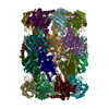
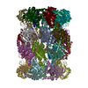
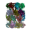
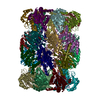
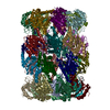
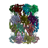
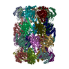
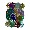
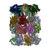
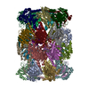
- PDB-3mi0: Crystal Structure of Mycobacterium Tuberculosis Proteasome at 2.2 A -
+ Open data

- Basic information
| Entry | Database: PDB / ID: 3mi0 | ||||||
|---|---|---|---|---|---|---|---|
| Title | Crystal Structure of Mycobacterium Tuberculosis Proteasome at 2.2 A | ||||||
 Components Components |
| ||||||
 Keywords Keywords |  HYDROLASE / Enzyme Inhibitors / Lactones / Proteasome Endopeptidase Complex / Mycobacterium tuberculosis HYDROLASE / Enzyme Inhibitors / Lactones / Proteasome Endopeptidase Complex / Mycobacterium tuberculosis | ||||||
| Function / homology |  Function and homology information Function and homology informationsymbiont-mediated perturbation of host defenses / zymogen binding / cell wall / proteasome endopeptidase complex / proteasome core complex, beta-subunit complex / proteasome core complex, alpha-subunit complex / threonine-type endopeptidase activity / proteolysis involved in protein catabolic process / peptidoglycan-based cell wall / proteasomal protein catabolic process ...symbiont-mediated perturbation of host defenses / zymogen binding / cell wall / proteasome endopeptidase complex / proteasome core complex, beta-subunit complex / proteasome core complex, alpha-subunit complex / threonine-type endopeptidase activity / proteolysis involved in protein catabolic process / peptidoglycan-based cell wall / proteasomal protein catabolic process / modification-dependent protein catabolic process / extracellular region / plasma membrane / cytoplasmSimilarity search - Function | ||||||
| Biological species |   Mycobacterium tuberculosis (bacteria) Mycobacterium tuberculosis (bacteria) | ||||||
| Method | X-RAY DIFFRACTION / SYNCHROTRON / MOLECULAR REPLACEMENT / Resolution: 2.2 Å | ||||||
 Authors Authors | Li, D. / Li, H. | ||||||
 Citation Citation | Journal: Embo J. / Year: 2010 Title: Structural basis for the assembly and gate closure mechanisms of the Mycobacterium tuberculosis 20S proteasome. Authors: Li, D. / Li, H. / Wang, T. / Pan, H. / Lin, G. / Li, H. | ||||||
| History |
|
- Structure visualization
| Structure viewer | Molecule: MolmilJmol/JSmol |
|---|
- Downloads & links
-Download
| PDBx/mmCIF format | 3mi0.cif.gz | 1.2 MB | Display | PDBx/mmCIF format |
|---|---|---|---|---|
| PDB format | pdb3mi0.ent.gz | 1006.5 KB | Display | PDB format |
| PDBx/mmJSON format | 3mi0.json.gz | Tree view | PDBx/mmJSON format | |
| Others |  Other downloads Other downloads |
-Validation report
| Arichive directory | https://data.pdbj.org/pub/pdb/validation_reports/mi/3mi0ftp://data.pdbj.org/pub/pdb/validation_reports/mi/3mi0 | HTTPS FTP |
|---|
-Related structure data
| Related structure data | 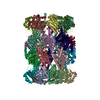 3mfeC  3mkaC 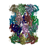 2fhgS S: Starting model for refinement C: citing same article ( |
|---|---|
| Similar structure data |


-Links
 PDBj
PDBj


- Assembly
| Deposited unit | 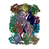
| ||||||||
|---|---|---|---|---|---|---|---|---|---|
| 1 |
| ||||||||
| Unit cell |
|
-Components
| #1: Protein | / Proteasome core protein prcA / 20S proteasome alpha subunit Mass: 26911.039 Da / Num. of mol.: 14 Source method: isolated from a genetically manipulated source Source: (gene. exp.) Mycobacterium tuberculosis (bacteria) / Strain: rv2109c / Gene: MT2169, prcA, Rv2109c / Plasmid: pACYCDuet / Production host: Escherichia coli (E. coli) / Strain (production host): BL21References: UniProt: O33244, UniProt: P9WHU1*PLUS, proteasome endopeptidase complex#2: Protein | / Proteasome core protein prcB / 20S proteasome beta subunitMass: 25274.264 Da / Num. of mol.: 14 Source method: isolated from a genetically manipulated source Source: (gene. exp.) Mycobacterium tuberculosis (bacteria) / Strain: rv2109c / Gene: MT2170, prcB, Rv2110c / Plasmid: pACYCDuet / Production host: Escherichia coli (E. coli) / Strain (production host): BL21References: UniProt: O33245, UniProt: P9WHT9*PLUS, proteasome endopeptidase complex#3: Chemical | ChemComp-DMF / Dimethylformamide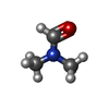  Mass: 73.094 Da / Num. of mol.: 123 / Source method: obtained synthetically / Formula: C3H7NO Mass: 73.094 Da / Num. of mol.: 123 / Source method: obtained synthetically / Formula: C3H7NO#4: Chemical | ChemComp-SA6 / ( 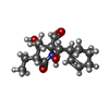  Mass: 281.347 Da / Num. of mol.: 14 / Source method: obtained synthetically / Formula: C15H23NO4 Mass: 281.347 Da / Num. of mol.: 14 / Source method: obtained synthetically / Formula: C15H23NO4#5: Water | ChemComp-HOH / | Water Mass: 18.015 Da / Num. of mol.: 2680 / Source method: isolated from a natural source / Formula: H2O Mass: 18.015 Da / Num. of mol.: 2680 / Source method: isolated from a natural source / Formula: H2O |
|---|
-Experimental details
-Experiment
| Experiment | Method: X-RAY DIFFRACTION / Number of used crystals: 1 |
|---|
- Sample preparation
| Crystal | Density Matthews: 2.48 Å3/Da / Density % sol: 50.43 % |
|---|---|
| Crystal grow | Temperature: 277 K / Method: vapor diffusion, sitting drop / pH: 5.7 Details: 13.2% PEG 6000, 60mM sodium citrate, pH 5.7, VAPOR DIFFUSION, SITTING DROP, temperature 277.0K |
-Data collection
| Diffraction | Mean temperature: 100 K |
|---|---|
| Diffraction source | Source: SYNCHROTRON / Site: NSLS  / Beamline: X29A / Wavelength: 1.0809 Å / Beamline: X29A / Wavelength: 1.0809 Å |
| Detector | Type: ADSC QUANTUM 4 / Detector: CCD / Date: Oct 28, 2008 |
| Radiation | Monochromator: Si 111 CHANNEL / Protocol: SINGLE WAVELENGTH / Monochromatic (M) / Laue (L): M / Scattering type: x-ray |
| Radiation wavelength | Wavelength: 1.0809 Å / Relative weight: 1 |
| Reflection | Resolution: 2.1→25 Å / Num. all: 378907 / Num. obs: 362039 / % possible obs: 94.3 % / Observed criterion σ(F): 0 / Observed criterion σ(I): 0 / Redundancy: 4.6 % / Biso Wilson estimate: 14 Å2 / Rmerge(I) obs: 0.117 / Net I/σ(I): 9.7 |
| Reflection shell | Resolution: 2.1→2.18 Å / Redundancy: 2.6 % / Rmerge(I) obs: 0.641 / Mean I/σ(I) obs: 1.4 / Num. unique all: 33082 / % possible all: 82.7 |
- Processing
| Software |
| ||||||||||||||||||||||||||||||||||||||||||||||||||||||||||||
|---|---|---|---|---|---|---|---|---|---|---|---|---|---|---|---|---|---|---|---|---|---|---|---|---|---|---|---|---|---|---|---|---|---|---|---|---|---|---|---|---|---|---|---|---|---|---|---|---|---|---|---|---|---|---|---|---|---|---|---|---|---|
| Refinement | Method to determine structure: MOLECULAR REPLACEMENT Starting model: PDB entry 2FHG Resolution: 2.2→24.95 Å / Rfactor Rfree error: 0.002 / Data cutoff high absF: 96363.28 / Data cutoff low absF: 0 / Isotropic thermal model: RESTRAINED / Cross valid method: THROUGHOUT / σ(F): 0 / Stereochemistry target values: Engh & Huber / Details: BULK SOLVENT MODEL USED
| ||||||||||||||||||||||||||||||||||||||||||||||||||||||||||||
| Solvent computation | Solvent model: FLAT MODEL / Bsol: 48.3711 Å2 / ksol: 0.35 e/Å3 | ||||||||||||||||||||||||||||||||||||||||||||||||||||||||||||
| Displacement parameters | Biso mean: 40.5 Å2
| ||||||||||||||||||||||||||||||||||||||||||||||||||||||||||||
| Refine analyze |
| ||||||||||||||||||||||||||||||||||||||||||||||||||||||||||||
| Refinement step | Cycle: LAST / Resolution: 2.2→24.95 Å
| ||||||||||||||||||||||||||||||||||||||||||||||||||||||||||||
| Refine LS restraints |
| ||||||||||||||||||||||||||||||||||||||||||||||||||||||||||||
| Refine LS restraints NCS | NCS model details: CONSTR | ||||||||||||||||||||||||||||||||||||||||||||||||||||||||||||
| LS refinement shell | Resolution: 2.2→2.28 Å / Rfactor Rfree error: 0.008 / Total num. of bins used: 10
| ||||||||||||||||||||||||||||||||||||||||||||||||||||||||||||
| Xplor file |
|
