 Movie
Movie Controller

[English] 日本語
 Yorodumi
- PDB-3mc3: Crystal structure of DsrE/DsrF-like family protein (NP_342589.1) ... -
+ Open data

- Basic information
| Entry | Database: PDB / ID: 3mc3 | ||||||
|---|---|---|---|---|---|---|---|
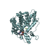
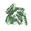
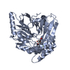
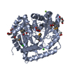
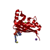
| Title | Crystal structure of DsrE/DsrF-like family protein (NP_342589.1) from SULFOLOBUS SOLFATARICUS at 1.49 A resolution | ||||||
 Components Components | DsrE/DsrF-like family protein | ||||||
 Keywords Keywords |  structural genomics / unknown function / Joint Center for Structural Genomics / JCSG / Protein Structure Initiative / PSI-2 structural genomics / unknown function / Joint Center for Structural Genomics / JCSG / Protein Structure Initiative / PSI-2 | ||||||
| Function / homology | Sulphur relay, DsrE/F-like / DsrE/DsrF-like family / DsrEFH-like / DsrEFH-like / Hypothetical Protein Ychn; Chain: A, / 3-Layer(aba) Sandwich / Alpha Beta / DrsE domain-containing protein Function and homology information Function and homology information | ||||||
| Biological species |   Sulfolobus solfataricus (archaea) Sulfolobus solfataricus (archaea) | ||||||
| Method | X-RAY DIFFRACTION / SYNCHROTRON / MAD / Resolution: 1.49 Å | ||||||
 Authors Authors | Joint Center for Structural Genomics (JCSG) | ||||||
 Citation Citation | Journal: To be published Title: Crystal structure of DsrE/DsrF-like family protein (NP_342589.1) from SULFOLOBUS SOLFATARICUS at 1.49 A resolution Authors: Joint Center for Structural Genomics (JCSG) | ||||||
| History |
|
- Structure visualization
| Structure viewer | Molecule: MolmilJmol/JSmol |
|---|
- Downloads & links
-Download
| PDBx/mmCIF format | 3mc3.cif.gz | 41 KB | Display | PDBx/mmCIF format |
|---|---|---|---|---|
| PDB format | pdb3mc3.ent.gz | 31.8 KB | Display | PDB format |
| PDBx/mmJSON format | 3mc3.json.gz | Tree view | PDBx/mmJSON format | |
| Others |  Other downloads Other downloads |
-Validation report
| Arichive directory | https://data.pdbj.org/pub/pdb/validation_reports/mc/3mc3ftp://data.pdbj.org/pub/pdb/validation_reports/mc/3mc3 | HTTPS FTP |
|---|
-Related structure data
| Similar structure data | |
|---|---|
| Other databases |


-Links
 PDBj
PDBj- Assembly
| Deposited unit | 
| |||||||||
|---|---|---|---|---|---|---|---|---|---|---|
| 1 | 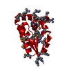
| |||||||||
| Unit cell |
| |||||||||
| Components on special symmetry positions |
|
-Components
| #1: Protein | Mass: 16019.342 Da / Num. of mol.: 1 Source method: isolated from a genetically manipulated source Source: (gene. exp.) Sulfolobus solfataricus (archaea) / Gene: SSO1125 / Plasmid: SpeedET / Production host:  Escherichia Coli (E. coli) / Strain (production host): HK100 / References: UniProt: Q97Z18 Escherichia Coli (E. coli) / Strain (production host): HK100 / References: UniProt: Q97Z18 |
|---|---|
| #2: Chemical | ChemComp-CL / Chloride  Mass: 35.453 Da / Num. of mol.: 1 / Source method: obtained synthetically / Formula: Cl Mass: 35.453 Da / Num. of mol.: 1 / Source method: obtained synthetically / Formula: Cl |
| #3: Water | ChemComp-HOH / Water Mass: 18.015 Da / Num. of mol.: 111 / Source method: isolated from a natural source / Formula: H2O Mass: 18.015 Da / Num. of mol.: 111 / Source method: isolated from a natural source / Formula: H2O |
| Sequence details | 1. THIS CONSTRUCT WAS EXPRESSED WITH AN N-TERMINAL PURIFICATION TAG MGSDKIHHHHHHENLYFQG. THE TAG ...1. THIS CONSTRUCT WAS EXPRESSED WITH AN N-TERMINAL PURIFICATI |
-Experimental details
-Experiment
| Experiment | Method: X-RAY DIFFRACTION / Number of used crystals: 1 |
|---|
- Sample preparation
| Crystal | Density Matthews: 2.61 Å3/Da / Density % sol: 52.8 % Description: THE STATISTICS REPORTED IN REMARK 200 WERE COMPUTED WITH XSCALE WITH FRIEDEL PAIRS KEPT SEPARATE. |
|---|---|
| Crystal grow | Temperature: 277 K / Method: vapor diffusion, sitting drop / pH: 6 Details: 5.0000% polyethylene glycol 3000, 40.0000% polyethylene glycol 400, 0.1M MES pH 6.0, NANODROP', VAPOR DIFFUSION, SITTING DROP, temperature 277K |
-Data collection
| Diffraction | Mean temperature: 100 K | |||||||||||||||||||||||||||||||||||||||||||||||||||||||||||||||||||||||||||||
|---|---|---|---|---|---|---|---|---|---|---|---|---|---|---|---|---|---|---|---|---|---|---|---|---|---|---|---|---|---|---|---|---|---|---|---|---|---|---|---|---|---|---|---|---|---|---|---|---|---|---|---|---|---|---|---|---|---|---|---|---|---|---|---|---|---|---|---|---|---|---|---|---|---|---|---|---|---|---|
| Diffraction source | Source: SYNCHROTRON / Site: SSRL  / Beamline: BL11-1 / Wavelength: 0.91837,0.97871,0.97831 / Beamline: BL11-1 / Wavelength: 0.91837,0.97871,0.97831 | |||||||||||||||||||||||||||||||||||||||||||||||||||||||||||||||||||||||||||||
| Detector | Type: MARMOSAIC 325 mm CCD / Detector: CCD / Date: Nov 15, 2008 / Details: Flat mirror (vertical focusing) | |||||||||||||||||||||||||||||||||||||||||||||||||||||||||||||||||||||||||||||
| Radiation | Monochromator: Single crystal Si(111) bent monochromator (horizontal focusing) Protocol: MAD / Monochromatic (M) / Laue (L): M / Scattering type: x-ray | |||||||||||||||||||||||||||||||||||||||||||||||||||||||||||||||||||||||||||||
| Radiation wavelength |
| |||||||||||||||||||||||||||||||||||||||||||||||||||||||||||||||||||||||||||||
| Reflection twin |
| |||||||||||||||||||||||||||||||||||||||||||||||||||||||||||||||||||||||||||||
| Reflection | Resolution: 1.49→26.832 Å / Num. obs: 26406 / % possible obs: 96.1 % / Observed criterion σ(I): -3 / Biso Wilson estimate: 17.255 Å2 / Rmerge(I) obs: 0.03 / Net I/σ(I): 13.48 | |||||||||||||||||||||||||||||||||||||||||||||||||||||||||||||||||||||||||||||
| Reflection shell |
|
-Phasing
| Phasing | Method: MAD |
|---|
- Processing
| Software |
| |||||||||||||||||||||||||||||||||||||||||||||||||||||||||||||||||||||||||||||||||||||
|---|---|---|---|---|---|---|---|---|---|---|---|---|---|---|---|---|---|---|---|---|---|---|---|---|---|---|---|---|---|---|---|---|---|---|---|---|---|---|---|---|---|---|---|---|---|---|---|---|---|---|---|---|---|---|---|---|---|---|---|---|---|---|---|---|---|---|---|---|---|---|---|---|---|---|---|---|---|---|---|---|---|---|---|---|---|---|
| Refinement | Method to determine structure: MAD / Resolution: 1.49→26.832 Å / Cor.coef. Fo:Fc: 0.981 / Cor.coef. Fo:Fc free: 0.973 / Occupancy max: 1 / Occupancy min: 0.15 / SU B: 1.693 / SU ML: 0.027 / TLS residual ADP flag: LIKELY RESIDUAL / Cross valid method: THROUGHOUT / σ(F): 0 / ESU R: 0.009 / ESU R Free: 0.01 / Stereochemistry target values: MAXIMUM LIKELIHOOD Details: 1. HYDROGENS HAVE BEEN ADDED IN THE RIDING POSITIONS. 2. ATOM RECORDS CONTAIN RESIDUAL B FACTORS ONLY. 3. A MET-INHIBITION PROTOCOL WAS USED FOR SELENOMETHIONINE INCORPORATION DURING PROTEIN ...Details: 1. HYDROGENS HAVE BEEN ADDED IN THE RIDING POSITIONS. 2. ATOM RECORDS CONTAIN RESIDUAL B FACTORS ONLY. 3. A MET-INHIBITION PROTOCOL WAS USED FOR SELENOMETHIONINE INCORPORATION DURING PROTEIN EXPRESSION. THE OCCUPANCY OF THE SE ATOMS IN THE MSE RESIDUES WAS REDUCED TO 0.75 FOR THE REDUCED SCATTERING POWER DUE TO PARTIAL S-MET INCORPORATION. 4. THE DATA IS TWINNED. THE FINAL TWIN FRACTION WAS REFINED TO 0.228 USING THE OPERATOR "K,H,-L". 5.THE PROTEIN WAS SUBJECTED TO REDUCTIVE METHYLATION PRIOR TO CRYSTALLIZATION. THE LYSINE RESIDUES HAVE BEEN MODELED AS N-DIMETHYL-LYSINE (MLY). 6. CYSTEINE 93 IS OXIDIZED AND IS MODELED AS S-OXY CYSTEINE (CSX). 7. CHLORIDE ION (CL) IS MODELED FROM THE PROTEIN BUFFER SOLUTION.
| |||||||||||||||||||||||||||||||||||||||||||||||||||||||||||||||||||||||||||||||||||||
| Solvent computation | Ion probe radii: 0.8 Å / Shrinkage radii: 0.8 Å / VDW probe radii: 1.4 Å / Solvent model: BABINET MODEL WITH MASK | |||||||||||||||||||||||||||||||||||||||||||||||||||||||||||||||||||||||||||||||||||||
| Displacement parameters | Biso max: 56.28 Å2 / Biso mean: 14.739 Å2 / Biso min: 2 Å2
| |||||||||||||||||||||||||||||||||||||||||||||||||||||||||||||||||||||||||||||||||||||
| Refinement step | Cycle: LAST / Resolution: 1.49→26.832 Å
| |||||||||||||||||||||||||||||||||||||||||||||||||||||||||||||||||||||||||||||||||||||
| Refine LS restraints |
| |||||||||||||||||||||||||||||||||||||||||||||||||||||||||||||||||||||||||||||||||||||
| LS refinement shell | Resolution: 1.49→1.528 Å / Total num. of bins used: 20
| |||||||||||||||||||||||||||||||||||||||||||||||||||||||||||||||||||||||||||||||||||||
| Refinement TLS params. | Method: refined / Origin x: 10.354 Å / Origin y: 10.474 Å / Origin z: 59.661 Å
|
