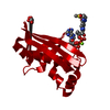
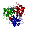
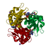
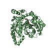
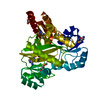
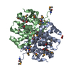
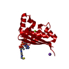
Mass: 14751.777 Da / Num. of mol.: 1 Source method: isolated from a genetically manipulated source Details: THE PROTEIN IS IN COMPLEX WITH THE COFACTOR COA / Source: (gene. exp.) STREPTOMYCES COELICOLOR (bacteria) / Plasmid: PET15B / Production host: ESCHERICHIA COLI (E. coli) / Strain (production host): BL21(DE3) References: UniProt: O86785, holo-[acyl-carrier-protein] synthase
Mass: 18.015 Da / Num. of mol.: 183 / Source method: isolated from a natural source / Formula: H2O
-
Experimental details
-
Experiment
Experiment
Method: X-RAY DIFFRACTION / Number of used crystals: 1
-
Sample preparation
Crystal
Density Matthews: 2.21 Å3/Da / Density % sol: 44.35 %
Crystal grow
pH: 6.5 Details: THE PROTEIN WAS DIALYSED IN 50 MM TRIS, 10 MM MGCL2, 10% GLYCEROL, PH 8. 2.5 MM COA WAS SOAKED INTO THE PROTEIN. THE PROTEIN WAS THEN CRYSTALLYZED IN 0.3M KSCN, 15% PEG 4K, 0.1M NACACODYLATE PH 6.5.
Type: MARRESEARCH / Detector: CCD / Date: Nov 10, 2006
Radiation
Protocol: SINGLE WAVELENGTH / Monochromatic (M) / Laue (L): M / Scattering type: x-ray
Radiation wavelength
Wavelength: 1.488 Å / Relative weight: 1
Reflection
Resolution: 1.62→50 Å / Num. obs: 16798 / % possible obs: 90.7 % / Redundancy: 6.1 % / Biso Wilson estimate: 18.6 Å2 / Rmerge(I) obs: 0.08 / Net I/σ(I): 18.88
Reflection shell
Resolution: 1.62→1.68 Å / Redundancy: 1.8 % / Rmerge(I) obs: 0.33 / Mean I/σ(I) obs: 2.15 / % possible all: 69.3
-
Processing
Software
Name
Version
Classification
REFMAC
5.2.0019
refinement
DENZO
datareduction
SCALEPACK
datascaling
Refinement
Method to determine structure: MOLECULAR REPLACEMENT Starting model: S. COELICOLOR APO- ACPS Resolution: 1.62→51.71 Å / Cor.coef. Fo:Fc: 0.956 / Cor.coef. Fo:Fc free: 0.943 / SU ML: 0.069 / Cross valid method: THROUGHOUT / ESU R: 0.109 / ESU R Free: 0.103 / Stereochemistry target values: MAXIMUM LIKELIHOOD Details: HYDROGENS HAVE BEEN ADDED IN THE RIDING POSITIONS. THE OCCUPANCY OF RELATIVELY DISORDERED PARTS OF THE SIDE CHAIN OF ARG15, GLU98 AND ASP111 WAS SET TO 0.5 TO GET RID OF NEGATIVE DENSITY.
Rfactor
Num. reflection
% reflection
Selection details
Rfree
0.215
771
5.1 %
RANDOM
Rwork
0.187
-
-
-
obs
0.189
14453
90.6 %
-
Solvent computation
Ion probe radii: 0.8 Å / Shrinkage radii: 0.8 Å / VDW probe radii: 1.4 Å / Solvent model: BABINET MODEL WITH MASK
 Movie
Movie Controller

 Yorodumi
 Open data
 Basic information
 Components
 Keywords
 TRANSFERASE / ACP /
  Function and homology information
 Authors
 Citation
 Structure visualization
 Downloads & links
 Other downloads


 PDBj
PDBj


 Assembly

 Mass: 24.305 Da / Num. of mol.: 1 / Source method: obtained synthetically / Formula: Mg
 Mass: 39.098 Da / Num. of mol.: 1 / Source method: obtained synthetically / Formula: K
 Mass: 767.534 Da / Num. of mol.: 1 / Source method: obtained synthetically / Formula: C21H36N7O16P3S
 Mass: 18.015 Da / Num. of mol.: 183 / Source method: isolated from a natural source / Formula: H2O
 Sample preparation
 / Beamline: PX10.1 / Wavelength: 1.488
  Processing