 Movie
Movie Controller

[English] 日本語
 Yorodumi
- PDB-2wdy: Crystal structure of the Streptomyces coelicolor D111A AcpS mutan... -
+ Open data

- Basic information
| Entry | Database: PDB / ID: 2wdy | ||||||
|---|---|---|---|---|---|---|---|
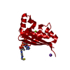
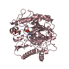
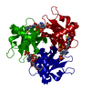
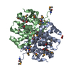
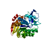
| Title | Crystal structure of the Streptomyces coelicolor D111A AcpS mutant in complex with cofactor CoA at 1.4 A | ||||||
 Components Components | HOLO-[ACYL-CARRIER-PROTEIN] SYNTHASE | ||||||
 Keywords Keywords |  TRANSFERASE / PHOSPHOPANTETHEINE ARM / FATTY ACID BIOSYNTHESIS / LIPID SYNTHESIS / POLYKETIDES TRANSFERASE / PHOSPHOPANTETHEINE ARM / FATTY ACID BIOSYNTHESIS / LIPID SYNTHESIS / POLYKETIDES | ||||||
| Function / homology |  Function and homology information Function and homology informationholo-[acyl-carrier-protein] synthase / holo-[acyl-carrier-protein] synthase activity / fatty acid biosynthetic process / magnesium ion binding / cytoplasmSimilarity search - Function | ||||||
| Biological species |  STREPTOMYCES COELICOLOR (bacteria) STREPTOMYCES COELICOLOR (bacteria) | ||||||
| Method | X-RAY DIFFRACTION / SYNCHROTRON / MOLECULAR REPLACEMENT / Resolution: 1.4 Å | ||||||
 Authors Authors | Dall'Aglio, P. / Arthur, C. / Crump, M.P. / Crosby, J. / Hadfield, A.T. | ||||||
 Citation Citation | Journal: Biochemistry / Year: 2011 Title: Analysis of Streptomyces Coelicolor Phosphopantetheinyl Transferase, Acps, Reveals the Basis for Relaxed Substrate Specificity. Authors: Dall'Aglio, P. / Arthur, C. / Williams, C. / Vasilakis, K. / Maple, H.J. / Crosby, J. / Crump, M.P. / Hadfield, A.T. | ||||||
| History |
|
- Structure visualization
| Structure viewer | Molecule: MolmilJmol/JSmol |
|---|
- Downloads & links
-Download
| PDBx/mmCIF format | 2wdy.cif.gz | 46.8 KB | Display | PDBx/mmCIF format |
|---|---|---|---|---|
| PDB format | pdb2wdy.ent.gz | 32.6 KB | Display | PDB format |
| PDBx/mmJSON format | 2wdy.json.gz | Tree view | PDBx/mmJSON format | |
| Others |  Other downloads Other downloads |
-Validation report
| Arichive directory | https://data.pdbj.org/pub/pdb/validation_reports/wd/2wdyftp://data.pdbj.org/pub/pdb/validation_reports/wd/2wdy | HTTPS FTP |
|---|
-Related structure data
| Related structure data |  2jbzSC  2jcaC 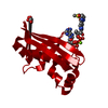 2wdoC  2wdsC C: citing same article ( S: Starting model for refinement |
|---|---|
| Similar structure data |

-Links
 PDBj
PDBj


- Assembly
| Deposited unit | 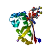
| |||||||||||||||||||||||||||
|---|---|---|---|---|---|---|---|---|---|---|---|---|---|---|---|---|---|---|---|---|---|---|---|---|---|---|---|---|
| 1 | 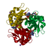
| |||||||||||||||||||||||||||
| Unit cell |
| |||||||||||||||||||||||||||
| Components on special symmetry positions |
|
-Components
| #1: Protein | Mass: 14707.768 Da / Num. of mol.: 1 / Mutation: YES Source method: isolated from a genetically manipulated source Details: MG AND COA ARE PRESENT IN THE ACTIVE SITE. INSPECTION OF THE ELECTRON DENSITY SUGGESTS COA BINDS IN 2 ALTERNATIVE CONFORMATIONS. Source: (gene. exp.) STREPTOMYCES COELICOLOR (bacteria) / Production host: ESCHERICHIA COLI (E. coli) / Strain (production host): BL21(DE3)References: UniProt: O86785, holo-[acyl-carrier-protein] synthase | ||||||||
|---|---|---|---|---|---|---|---|---|---|
| #2: Chemical |   Mass: 24.305 Da / Num. of mol.: 2 / Source method: obtained synthetically / Formula: Mg Mass: 24.305 Da / Num. of mol.: 2 / Source method: obtained synthetically / Formula: Mg#3: Chemical | ChemComp-NA / |   Mass: 22.990 Da / Num. of mol.: 1 / Source method: obtained synthetically / Formula: Na Mass: 22.990 Da / Num. of mol.: 1 / Source method: obtained synthetically / Formula: Na#4: Chemical | ChemComp-COA / | Coenzyme A  Mass: 767.534 Da / Num. of mol.: 1 / Source method: obtained synthetically / Formula: C21H36N7O16P3S Mass: 767.534 Da / Num. of mol.: 1 / Source method: obtained synthetically / Formula: C21H36N7O16P3S#5: Water | ChemComp-HOH / | Water Mass: 18.015 Da / Num. of mol.: 207 / Source method: isolated from a natural source / Formula: H2O Mass: 18.015 Da / Num. of mol.: 207 / Source method: isolated from a natural source / Formula: H2OCompound details | ENGINEERED | |
-Experimental details
-Experiment
| Experiment | Method: X-RAY DIFFRACTION / Number of used crystals: 1 |
|---|
- Sample preparation
| Crystal | Density Matthews: 2.2 Å3/Da / Density % sol: 44 % / Description: NONE |
|---|---|
| Crystal grow | pH: 6.5 Details: 0.2 M POTASSIUM THIOCYANATE, 0.1 M SODIUM CACODYLATE PH 6.5, 8% PEG 20K PLUS 8% PEG 550 MME, 15% GLYCEROL WAS THEN ADDED AS CRYO |
-Data collection
| Diffraction | Mean temperature: 100 K |
|---|---|
| Diffraction source | Source: SYNCHROTRON / Site: Diamond  / Beamline: I02 / Wavelength: 0.953 / Beamline: I02 / Wavelength: 0.953 |
| Detector | Type: ADSC CCD / Detector: CCD / Date: Feb 11, 2008 |
| Radiation | Protocol: SINGLE WAVELENGTH / Monochromatic (M) / Laue (L): M / Scattering type: x-ray |
| Radiation wavelength | Wavelength: 0.953 Å / Relative weight: 1 |
| Reflection | Resolution: 1.4→50 Å / Num. obs: 25707 / % possible obs: 99.6 % / Observed criterion σ(I): 2 / Redundancy: 7.9 % / Rmerge(I) obs: 0.05 / Net I/σ(I): 34.7 |
| Reflection shell | Resolution: 1.4→1.45 Å / Redundancy: 2.9 % / Rmerge(I) obs: 0.51 / Mean I/σ(I) obs: 1.5 / % possible all: 96.4 |
- Processing
| Software |
| ||||||||||||||||||||||||||||||||||||||||||||||||||||||||||||||||||||||||||||||||||||||||||||||||||||||||||||||||||||||||||||||||||||||||||||||||||||||||||||||||||||||||||||||||||||||
|---|---|---|---|---|---|---|---|---|---|---|---|---|---|---|---|---|---|---|---|---|---|---|---|---|---|---|---|---|---|---|---|---|---|---|---|---|---|---|---|---|---|---|---|---|---|---|---|---|---|---|---|---|---|---|---|---|---|---|---|---|---|---|---|---|---|---|---|---|---|---|---|---|---|---|---|---|---|---|---|---|---|---|---|---|---|---|---|---|---|---|---|---|---|---|---|---|---|---|---|---|---|---|---|---|---|---|---|---|---|---|---|---|---|---|---|---|---|---|---|---|---|---|---|---|---|---|---|---|---|---|---|---|---|---|---|---|---|---|---|---|---|---|---|---|---|---|---|---|---|---|---|---|---|---|---|---|---|---|---|---|---|---|---|---|---|---|---|---|---|---|---|---|---|---|---|---|---|---|---|---|---|---|---|
| Refinement | Method to determine structure: MOLECULAR REPLACEMENT Starting model: PDB ENTRY 2JBZ Resolution: 1.4→51.5 Å / Cor.coef. Fo:Fc: 0.964 / Cor.coef. Fo:Fc free: 0.948 / SU B: 1.096 / SU ML: 0.045 / Cross valid method: THROUGHOUT / ESU R: 0.069 / ESU R Free: 0.07 / Stereochemistry target values: MAXIMUM LIKELIHOOD Details: HYDROGENS HAVE BEEN ADDED IN THE RIDING POSITIONS. COA WAS BOUND IN TWO ALTERNATIVE CONFORMATIONS. WHILST THE ELECTRON DENSITY FOR THE ADENINE RINGS OF BOTH COAS, INCLUDING THE PHOSPHATES, ...Details: HYDROGENS HAVE BEEN ADDED IN THE RIDING POSITIONS. COA WAS BOUND IN TWO ALTERNATIVE CONFORMATIONS. WHILST THE ELECTRON DENSITY FOR THE ADENINE RINGS OF BOTH COAS, INCLUDING THE PHOSPHATES, AND THEIR RELATED MAGNESIUM IONS WAS VERY CLEAR, THE DENSITY FOR THE PHOSPHOPANTETHEINE ARMS WAS NOT WELL DEFINED
| ||||||||||||||||||||||||||||||||||||||||||||||||||||||||||||||||||||||||||||||||||||||||||||||||||||||||||||||||||||||||||||||||||||||||||||||||||||||||||||||||||||||||||||||||||||||
| Solvent computation | Ion probe radii: 0.8 Å / Shrinkage radii: 0.8 Å / VDW probe radii: 1.4 Å / Solvent model: BABINET MODEL WITH MASK | ||||||||||||||||||||||||||||||||||||||||||||||||||||||||||||||||||||||||||||||||||||||||||||||||||||||||||||||||||||||||||||||||||||||||||||||||||||||||||||||||||||||||||||||||||||||
| Displacement parameters | Biso mean: 21.166 Å2 | ||||||||||||||||||||||||||||||||||||||||||||||||||||||||||||||||||||||||||||||||||||||||||||||||||||||||||||||||||||||||||||||||||||||||||||||||||||||||||||||||||||||||||||||||||||||
| Refinement step | Cycle: LAST / Resolution: 1.4→51.5 Å
| ||||||||||||||||||||||||||||||||||||||||||||||||||||||||||||||||||||||||||||||||||||||||||||||||||||||||||||||||||||||||||||||||||||||||||||||||||||||||||||||||||||||||||||||||||||||
| Refine LS restraints |
|