 Movie
Movie Controller

[English] 日本語
 Yorodumi
- PDB-3ma5: Crystal structure of the tetratricopeptide repeat domain protein ... -
+ Open data

- Basic information
| Entry | Database: PDB / ID: 3ma5 | ||||||
|---|---|---|---|---|---|---|---|
| Title | Crystal structure of the tetratricopeptide repeat domain protein Q2S6C5_SALRD from Salinibacter ruber. Northeast Structural Genomics Consortium Target SrR115c. | ||||||
 Components Components | Tetratricopeptide repeat domain protein Tetratricopeptide repeat Tetratricopeptide repeat | ||||||
 Keywords Keywords | structural genomics / unknown function / Tetratricopeptide repeat domain protein / NESG / PSI-2 / Protein Structure Initiative / Northeast Structural Genomics Consortium | ||||||
| Function / homology |  Function and homology informationTetratricopeptide repeat / Tetratricopeptide repeat domain / Tetratricopeptide repeat / TPR repeat profile. / Tetratricopeptide repeats / Tetratricopeptide repeat / Serine Threonine Protein Phosphatase 5, Tetratricopeptide repeat / Alpha Horseshoe / Tetratricopeptide-like helical domain superfamily / Mainly Alpha Function and homology informationTetratricopeptide repeat / Tetratricopeptide repeat domain / Tetratricopeptide repeat / TPR repeat profile. / Tetratricopeptide repeats / Tetratricopeptide repeat / Serine Threonine Protein Phosphatase 5, Tetratricopeptide repeat / Alpha Horseshoe / Tetratricopeptide-like helical domain superfamily / Mainly AlphaSimilarity search - Domain/homology | ||||||
| Biological species |   Salinibacter ruber (bacteria) Salinibacter ruber (bacteria) | ||||||
| Method | X-RAY DIFFRACTION / SYNCHROTRON / MOLECULAR REPLACEMENT / Resolution: 2.8 Å | ||||||
 Authors Authors | Vorobiev, S. / Neely, H. / Seetharaman, J. / Wang, H. / Foote, E.L. / Ciccosanti, C. / Mao, L. / Xiao, R. / Acton, T.B. / Montelione, G.T. ...Vorobiev, S. / Neely, H. / Seetharaman, J. / Wang, H. / Foote, E.L. / Ciccosanti, C. / Mao, L. / Xiao, R. / Acton, T.B. / Montelione, G.T. / Hunt, J.F. / Tong, L. / Northeast Structural Genomics Consortium (NESG) | ||||||
 Citation Citation | Journal: To be Published Title: Crystal structure of the tetratricopeptide repeat domain protein Q2S6C5_SALRD from Salinibacter ruber. Authors: Vorobiev, S. / Neely, H. / Seetharaman, J. / Wang, H. / Foote, E.L. / Ciccosanti, C. / Mao, L. / Xiao, R. / Acton, T.B. / Montelione, G.T. / Hunt, J.F. / Tong, L. | ||||||
| History |
|
- Structure visualization
| Structure viewer | Molecule: MolmilJmol/JSmol |
|---|
- Downloads & links
-Download
| PDBx/mmCIF format | 3ma5.cif.gz | 78.1 KB | Display | PDBx/mmCIF format |
|---|---|---|---|---|
| PDB format | pdb3ma5.ent.gz | 59.8 KB | Display | PDB format |
| PDBx/mmJSON format | 3ma5.json.gz | Tree view | PDBx/mmJSON format | |
| Others |  Other downloads Other downloads |
-Validation report
| Arichive directory | https://data.pdbj.org/pub/pdb/validation_reports/ma/3ma5ftp://data.pdbj.org/pub/pdb/validation_reports/ma/3ma5 | HTTPS FTP |
|---|
-Related structure data
| Related structure data |  2kclS S: Starting model for refinement |
|---|---|
| Similar structure data | |
| Other databases |


-Links
 PDBj
PDBj


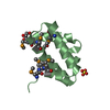
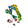
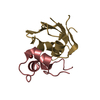
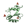
- Assembly
| Deposited unit | 
| ||||||||
|---|---|---|---|---|---|---|---|---|---|
| 1 | 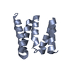
| ||||||||
| 2 | 
| ||||||||
| 3 | 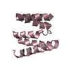
| ||||||||
| 4 | 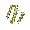
| ||||||||
| Unit cell |
| ||||||||
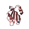
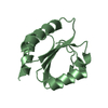
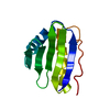
| Details | Dimer according to gel filtration |
-Components
| #1: Protein | Tetratricopeptide repeat Mass: 11618.613 Da / Num. of mol.: 4 Source method: isolated from a genetically manipulated source Source: (gene. exp.) Salinibacter ruber (bacteria) / Strain: HAMAP DSM 13855 / Gene: SRU_0103 / Plasmid: pET 21-23C / Production host: Escherichia coli (E. coli) / Strain (production host): BL21(DE3) +Magic / References: UniProt: Q2S6C5#2: Water | ChemComp-HOH / | Water Mass: 18.015 Da / Num. of mol.: 8 / Source method: isolated from a natural source / Formula: H2O Mass: 18.015 Da / Num. of mol.: 8 / Source method: isolated from a natural source / Formula: H2O |
|---|
-Experimental details
-Experiment
| Experiment | Method: X-RAY DIFFRACTION / Number of used crystals: 1 |
|---|
- Sample preparation
| Crystal | Density Matthews: 2.51 Å3/Da / Density % sol: 51.09 % |
|---|---|
| Crystal grow | Temperature: 291 K / Method: microbatch under paraffin oil / pH: 7 Details: 45-60% PEG 400, 0.03-0.05M potassium chloride, 0.1M Bis-Tris, pH 7.0, microbatch under Paraffin oil, temperature 291K |
-Data collection
| Diffraction source | Source: SYNCHROTRON / Site: NSLS  / Beamline: X4C / Wavelength: 0.97869 Å / Beamline: X4C / Wavelength: 0.97869 Å |
|---|---|
| Detector | Type: MAR CCD 165 mm / Detector: CCD / Date: Mar 15, 2010 |
| Radiation | Protocol: SINGLE WAVELENGTH / Monochromatic (M) / Laue (L): M / Scattering type: x-ray |
| Radiation wavelength | Wavelength: 0.97869 Å / Relative weight: 1 |
| Reflection | Resolution: 2.8→50 Å / Num. all: 22394 / Num. obs: 22372 / % possible obs: 99.9 % / Observed criterion σ(F): 0 / Observed criterion σ(I): 0 / Redundancy: 21.5 % / Rmerge(I) obs: 0.076 / Net I/σ(I): 59.1 |
| Reflection shell | Resolution: 2.8→2.9 Å / Redundancy: 22 % / Rmerge(I) obs: 0.941 / Mean I/σ(I) obs: 3.47 / Num. unique all: 2244 / % possible all: 100 |
- Processing
| Software |
| |||||||||||||||||||||||||||||||||||||||||||||||||||||||||||||||
|---|---|---|---|---|---|---|---|---|---|---|---|---|---|---|---|---|---|---|---|---|---|---|---|---|---|---|---|---|---|---|---|---|---|---|---|---|---|---|---|---|---|---|---|---|---|---|---|---|---|---|---|---|---|---|---|---|---|---|---|---|---|---|---|---|
| Refinement | Method to determine structure: MOLECULAR REPLACEMENT Starting model: PDB ENTRY 2KCL Resolution: 2.8→44.885 Å / SU ML: 0.42 / Cross valid method: THROUGHOUT / σ(F): 1.25 / Stereochemistry target values: ML Details: There are four molecules (two dimers) in AU. The fourth molecule (D) has very bad defined electron density and was not found by MR. Based on the partial electron density and dimer structure ...Details: There are four molecules (two dimers) in AU. The fourth molecule (D) has very bad defined electron density and was not found by MR. Based on the partial electron density and dimer structure C-terminal part (78-145) of molecule D was built; however B-factor for D molecule is higher than for A-C molecules.
| |||||||||||||||||||||||||||||||||||||||||||||||||||||||||||||||
| Solvent computation | Shrinkage radii: 0.9 Å / VDW probe radii: 1.11 Å / Solvent model: FLAT BULK SOLVENT MODEL / Bsol: 72.224 Å2 / ksol: 0.333 e/Å3 | |||||||||||||||||||||||||||||||||||||||||||||||||||||||||||||||
| Refinement step | Cycle: LAST / Resolution: 2.8→44.885 Å
| |||||||||||||||||||||||||||||||||||||||||||||||||||||||||||||||
| Refine LS restraints |
| |||||||||||||||||||||||||||||||||||||||||||||||||||||||||||||||
| LS refinement shell |
|
