 Movie
Movie Controller

[English] 日本語
 Yorodumi
- PDB-3m8a: Crystal Structure of Swine Flu Virus NS1 N-Terminal RNA Binding D... -
+ Open data

- Basic information
| Entry | Database: PDB / ID: 3m8a | ||||||
|---|---|---|---|---|---|---|---|
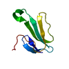
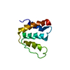
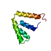
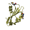
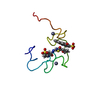
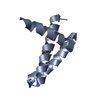
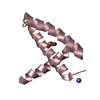
| Title | Crystal Structure of Swine Flu Virus NS1 N-Terminal RNA Binding Domain from H1N1 Influenza A/California/07/2009 | ||||||
 Components Components | Nonstructural protein 1 | ||||||
 Keywords Keywords |  VIRAL PROTEIN / NS1 / N-terminal RNA binding Domain / Influenza A virus / H1N1 / Host cytoplasm / Host nucleus / Host-virus interaction / Interferon antiviral system evasion / RNA-binding / Center for Structural Genomics of Infectious Diseases / CSGID VIRAL PROTEIN / NS1 / N-terminal RNA binding Domain / Influenza A virus / H1N1 / Host cytoplasm / Host nucleus / Host-virus interaction / Interferon antiviral system evasion / RNA-binding / Center for Structural Genomics of Infectious Diseases / CSGID | ||||||
| Function / homology |  Function and homology information Function and homology informationsymbiont-mediated suppression of host mRNA processing / symbiont-mediated suppression of host PKR/eIFalpha signaling / protein serine/threonine kinase inhibitor activity / symbiont-mediated suppression of host cytoplasmic pattern recognition receptor signaling pathway via inhibition of RIG-I activity / host cell cytoplasm / symbiont-mediated suppression of host type I interferon-mediated signaling pathway / host cell nucleus / RNA bindingSimilarity search - Function | ||||||
| Biological species |   Influenza A virus Influenza A virus | ||||||
| Method | X-RAY DIFFRACTION / SYNCHROTRON / MOLECULAR REPLACEMENT / Resolution: 2.1 Å | ||||||
 Authors Authors | Brunzelle, J.S. / Wawrzak, Z. / Skarina, T. / Savchenko, A. / Anderson, W.F. / Center for Structural Genomics of Infectious Diseases (CSGID) | ||||||
 Citation Citation | Journal: To be Published Title: Crystal Structure of Swine Flu Virus NS1 N-Terminal RNA Binding Domain from H1N1 Influenza A/California/07/2009 Authors: Brunzelle, J.S. / Wawrzak, Z. / Skarina, T. / Savchenko, A. / Anderson, W.F. / Center for Structural Genomics of Infectious Diseases (CSGID) | ||||||
| History |
|
- Structure visualization
| Structure viewer | Molecule: MolmilJmol/JSmol |
|---|
- Downloads & links
-Download
| PDBx/mmCIF format | 3m8a.cif.gz | 209.5 KB | Display | PDBx/mmCIF format |
|---|---|---|---|---|
| PDB format | pdb3m8a.ent.gz | 169.3 KB | Display | PDB format |
| PDBx/mmJSON format | 3m8a.json.gz | Tree view | PDBx/mmJSON format | |
| Others |  Other downloads Other downloads |
-Validation report
| Arichive directory | https://data.pdbj.org/pub/pdb/validation_reports/m8/3m8aftp://data.pdbj.org/pub/pdb/validation_reports/m8/3m8a | HTTPS FTP |
|---|
-Related structure data
| Related structure data | 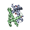 2z0aS S: Starting model for refinement |
|---|---|
| Similar structure data | |
| Other databases |


-Links
 PDBj
PDBj- Assembly


-Components
| #1: Protein | Mass: 8653.802 Da / Num. of mol.: 12 / Fragment: N-terminal RNA Binding Domain (UNP residues 1-73) Source method: isolated from a genetically manipulated source Source: (gene. exp.) Influenza A virus / Strain: A/CALIFORNIA/07/2009(H1N1) / References: UniProt: C3W611#2: Chemical | ChemComp-ACT / Acetate  Mass: 59.044 Da / Num. of mol.: 4 / Source method: obtained synthetically / Formula: C2H3O2 Mass: 59.044 Da / Num. of mol.: 4 / Source method: obtained synthetically / Formula: C2H3O2#3: Chemical | ChemComp-MLI / Malonic acid  Mass: 102.046 Da / Num. of mol.: 6 / Source method: obtained synthetically / Formula: C3H2O4 Mass: 102.046 Da / Num. of mol.: 6 / Source method: obtained synthetically / Formula: C3H2O4#4: Chemical | ChemComp-NA /   Mass: 22.990 Da / Num. of mol.: 4 / Source method: obtained synthetically / Formula: Na Mass: 22.990 Da / Num. of mol.: 4 / Source method: obtained synthetically / Formula: Na#5: Water | ChemComp-HOH / | Water Mass: 18.015 Da / Num. of mol.: 980 / Source method: isolated from a natural source / Formula: H2O Mass: 18.015 Da / Num. of mol.: 980 / Source method: isolated from a natural source / Formula: H2O |
|---|
-Experimental details
-Experiment
| Experiment | Method: X-RAY DIFFRACTION / Number of used crystals: 1 |
|---|
- Sample preparation
| Crystal | Density Matthews: 3.19 Å3/Da / Density % sol: 61.43 % |
|---|---|
| Crystal grow | Temperature: 298 K / Method: vapor diffusion, sitting drop / pH: 7 Details: Tacsimate 35%, ph 7, KCl 0.1M, MgCl2 0.1M, 0.3M NDSB211, TCEP 2mM, VAPOR DIFFUSION, SITTING DROP, temperature 298K |
-Data collection
| Diffraction | Mean temperature: 100 K |
|---|---|
| Diffraction source | Source: SYNCHROTRON / Site: APS  / Beamline: 21-ID-F / Wavelength: 0.97872 Å / Beamline: 21-ID-F / Wavelength: 0.97872 Å |
| Detector | Type: RAYONIX MX-225 / Detector: CCD / Date: Feb 4, 2010 / Details: Be Lenes |
| Radiation | Monochromator: Single Crystal Diamond / Protocol: SINGLE WAVELENGTH / Monochromatic (M) / Laue (L): M / Scattering type: x-ray |
| Radiation wavelength | Wavelength: 0.97872 Å / Relative weight: 1 |
| Reflection | Resolution: 2.1→30 Å / Num. all: 75867 / Num. obs: 75867 / % possible obs: 99.9 % / Observed criterion σ(F): -3 / Observed criterion σ(I): -3 / Redundancy: 6.4 % / Biso Wilson estimate: 28.36 Å2 / Rmerge(I) obs: 0.094 / Net I/σ(I): 18.9 |
| Reflection shell | Resolution: 2.1→2.14 Å / Redundancy: 6.2 % / Rmerge(I) obs: 0.511 / Mean I/σ(I) obs: 3.5 / % possible all: 99.8 |
- Processing
| Software |
| ||||||||||||||||||||||||||||||||||||||||||||||||||||||||||||||||||||||||||||||||||||||||||||||||||||||||||||||||||
|---|---|---|---|---|---|---|---|---|---|---|---|---|---|---|---|---|---|---|---|---|---|---|---|---|---|---|---|---|---|---|---|---|---|---|---|---|---|---|---|---|---|---|---|---|---|---|---|---|---|---|---|---|---|---|---|---|---|---|---|---|---|---|---|---|---|---|---|---|---|---|---|---|---|---|---|---|---|---|---|---|---|---|---|---|---|---|---|---|---|---|---|---|---|---|---|---|---|---|---|---|---|---|---|---|---|---|---|---|---|---|---|---|---|---|---|
| Refinement | Method to determine structure: MOLECULAR REPLACEMENT Starting model: PDB ENTRY 2Z0A Resolution: 2.1→29.25 Å / Cor.coef. Fo:Fc: 0.9429 / Cor.coef. Fo:Fc free: 0.9229 / Isotropic thermal model: Isotropic / Cross valid method: THROUGHOUT / σ(F): 0 / Stereochemistry target values: Engh & Huber
| ||||||||||||||||||||||||||||||||||||||||||||||||||||||||||||||||||||||||||||||||||||||||||||||||||||||||||||||||||
| Displacement parameters | Biso mean: 31.92 Å2
| ||||||||||||||||||||||||||||||||||||||||||||||||||||||||||||||||||||||||||||||||||||||||||||||||||||||||||||||||||
| Refine analyze | Luzzati coordinate error obs: 0.216 Å | ||||||||||||||||||||||||||||||||||||||||||||||||||||||||||||||||||||||||||||||||||||||||||||||||||||||||||||||||||
| Refinement step | Cycle: LAST / Resolution: 2.1→29.25 Å
| ||||||||||||||||||||||||||||||||||||||||||||||||||||||||||||||||||||||||||||||||||||||||||||||||||||||||||||||||||
| Refine LS restraints |
| ||||||||||||||||||||||||||||||||||||||||||||||||||||||||||||||||||||||||||||||||||||||||||||||||||||||||||||||||||
| LS refinement shell | Resolution: 2.1→2.15 Å / Total num. of bins used: 20
|
