 Movie
Movie Controller

[English] 日本語
 Yorodumi
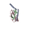
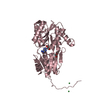
- PDB-3m5r: Crystal Structure of Swine Flu Virus NS1 Effector Domain from H1N... -
+ Open data

- Basic information
| Entry | Database: PDB / ID: 3m5r | ||||||
|---|---|---|---|---|---|---|---|
| Title | Crystal Structure of Swine Flu Virus NS1 Effector Domain from H1N1 Influenza A/California/07/2009 | ||||||
 Components Components | Nonstructural protein 1 | ||||||
 Keywords Keywords |  VIRAL PROTEIN / Swine Flu Virus / Influenza A H1N1 Subtype / Nonstructural Protein 1 / Effector domain / Viral immune evasion / Structural Genomics / NIAID / Center for Structural Genomics of Infectious Diseases / CSGID VIRAL PROTEIN / Swine Flu Virus / Influenza A H1N1 Subtype / Nonstructural Protein 1 / Effector domain / Viral immune evasion / Structural Genomics / NIAID / Center for Structural Genomics of Infectious Diseases / CSGID | ||||||
| Function / homology |  Function and homology information Function and homology informationsymbiont-mediated suppression of host mRNA processing / symbiont-mediated suppression of host PKR/eIFalpha signaling / protein serine/threonine kinase inhibitor activity / symbiont-mediated suppression of host cytoplasmic pattern recognition receptor signaling pathway via inhibition of RIG-I activity / host cell cytoplasm / symbiont-mediated suppression of host type I interferon-mediated signaling pathway / host cell nucleus / RNA bindingSimilarity search - Function | ||||||
| Biological species |   Influenza A virus Influenza A virus | ||||||
| Method | X-RAY DIFFRACTION / SYNCHROTRON / MOLECULAR REPLACEMENT / molecular replacement / Resolution: 2 Å | ||||||
 Authors Authors | Fremont, D.H. / Yu, Y.Y.L. / Nelson, C.A. / Center for Structural Genomics of Infectious Diseases (CSGID) | ||||||
 Citation Citation | Journal: To be Published Title: Crystal Structure of Swine Flu Virus NS1 Effector Domain from H1N1 Influenza A/California/07/2009 Authors: Fremont, D.H. / Yu, Y.Y.L. / Nelson, C.A. / Center for Structural Genomics of Infectious Diseases (CSGID) | ||||||
| History |
|
- Structure visualization
| Structure viewer | Molecule: MolmilJmol/JSmol |
|---|
- Downloads & links
-Download
| PDBx/mmCIF format | 3m5r.cif.gz | 159.3 KB | Display | PDBx/mmCIF format |
|---|---|---|---|---|
| PDB format | pdb3m5r.ent.gz | 126.3 KB | Display | PDB format |
| PDBx/mmJSON format | 3m5r.json.gz | Tree view | PDBx/mmJSON format | |
| Others |  Other downloads Other downloads |
-Validation report
| Arichive directory | https://data.pdbj.org/pub/pdb/validation_reports/m5/3m5rftp://data.pdbj.org/pub/pdb/validation_reports/m5/3m5r | HTTPS FTP |
|---|
-Related structure data
| Related structure data |  3f5tS S: Starting model for refinement |
|---|---|
| Similar structure data | |
| Other databases |


-Links
 PDBj
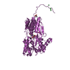
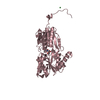
PDBj- Assembly
| Deposited unit | 
| ||||||||
|---|---|---|---|---|---|---|---|---|---|
| 1 | 
| ||||||||
| 2 | 
| ||||||||
| 3 | 
| ||||||||
| Unit cell |
|
-Components
| #1: Protein | Mass: 15038.358 Da / Num. of mol.: 6 / Fragment: C-TERMINAL EFFECTOR DOMAIN Source method: isolated from a genetically manipulated source Source: (gene. exp.) Influenza A virus / Strain: A/California/07/2009 (H1N1) / Gene: NS1 / Plasmid: pET28A(+) / Production host:  Escherichia coli (E. coli) / Strain (production host): BL21(DE3)RIL / References: UniProt: D2Y6Z6, UniProt: C3W611*PLUS Escherichia coli (E. coli) / Strain (production host): BL21(DE3)RIL / References: UniProt: D2Y6Z6, UniProt: C3W611*PLUS#2: Water | ChemComp-HOH / | Water Mass: 18.015 Da / Num. of mol.: 288 / Source method: isolated from a natural source / Formula: H2O Mass: 18.015 Da / Num. of mol.: 288 / Source method: isolated from a natural source / Formula: H2O |
|---|
-Experimental details
-Experiment
| Experiment | Method: X-RAY DIFFRACTION / Number of used crystals: 1 |
|---|
- Sample preparation
| Crystal | Density Matthews: 2.62 Å3/Da / Density % sol: 52.98 % |
|---|---|
| Crystal grow | Temperature: 293 K / Method: vapor diffusion, hanging drop / pH: 7.4 Details: 1.7 M NaCl, 10% Ethanol and 0.1 M hexamine cobalt (III) chloride, pH 7.4, VAPOR DIFFUSION, HANGING DROP, temperature 293K |
-Data collection
| Diffraction | Mean temperature: 100 K | ||||||||||||||||||||||||||||||||||||||||||||||||||||||||||||||||||
|---|---|---|---|---|---|---|---|---|---|---|---|---|---|---|---|---|---|---|---|---|---|---|---|---|---|---|---|---|---|---|---|---|---|---|---|---|---|---|---|---|---|---|---|---|---|---|---|---|---|---|---|---|---|---|---|---|---|---|---|---|---|---|---|---|---|---|---|
| Diffraction source | Source: SYNCHROTRON / Site: ALS  / Beamline: 4.2.2 / Wavelength: 1 Å / Beamline: 4.2.2 / Wavelength: 1 Å | ||||||||||||||||||||||||||||||||||||||||||||||||||||||||||||||||||
| Detector | Type: NOIR-1 / Detector: CCD / Date: Dec 18, 2009 | ||||||||||||||||||||||||||||||||||||||||||||||||||||||||||||||||||
| Radiation | Protocol: SINGLE WAVELENGTH / Monochromatic (M) / Laue (L): M / Scattering type: x-ray | ||||||||||||||||||||||||||||||||||||||||||||||||||||||||||||||||||
| Radiation wavelength | Wavelength: 1 Å / Relative weight: 1 | ||||||||||||||||||||||||||||||||||||||||||||||||||||||||||||||||||
| Reflection | Resolution: 2→100 Å / Num. obs: 61983 / % possible obs: 100 % / Redundancy: 3.8 % / Rmerge(I) obs: 0.061 / Χ2: 1.009 / Net I/σ(I): 18 | ||||||||||||||||||||||||||||||||||||||||||||||||||||||||||||||||||
| Reflection shell |
|
-Phasing
| Phasing | Method: molecular replacement |
|---|
- Processing
| Software |
| |||||||||||||||||||||||||||||||||||||||||||||||||||||||||||||||||||||||||||||
|---|---|---|---|---|---|---|---|---|---|---|---|---|---|---|---|---|---|---|---|---|---|---|---|---|---|---|---|---|---|---|---|---|---|---|---|---|---|---|---|---|---|---|---|---|---|---|---|---|---|---|---|---|---|---|---|---|---|---|---|---|---|---|---|---|---|---|---|---|---|---|---|---|---|---|---|---|---|---|
| Refinement | Method to determine structure: MOLECULAR REPLACEMENT Starting model: PDB ENTRY 3F5T Resolution: 2→50 Å / Occupancy max: 1 / Occupancy min: 1 / FOM work R set: 0.803 / SU ML: 0.3 / Cross valid method: THROUGHOUT / σ(F): 0 / Stereochemistry target values: ML
| |||||||||||||||||||||||||||||||||||||||||||||||||||||||||||||||||||||||||||||
| Solvent computation | Shrinkage radii: 0.9 Å / VDW probe radii: 1.11 Å / Solvent model: FLAT BULK SOLVENT MODEL / Bsol: 60.955 Å2 / ksol: 0.4 e/Å3 | |||||||||||||||||||||||||||||||||||||||||||||||||||||||||||||||||||||||||||||
| Displacement parameters | Biso max: 91.98 Å2 / Biso mean: 40.529 Å2 / Biso min: 18.33 Å2
| |||||||||||||||||||||||||||||||||||||||||||||||||||||||||||||||||||||||||||||
| Refinement step | Cycle: LAST / Resolution: 2→50 Å
| |||||||||||||||||||||||||||||||||||||||||||||||||||||||||||||||||||||||||||||
| Refine LS restraints |
| |||||||||||||||||||||||||||||||||||||||||||||||||||||||||||||||||||||||||||||
| LS refinement shell | Refine-ID: X-RAY DIFFRACTION / Total num. of bins used: 10
|
