 Movie
Movie Controller


+ Open data

- Basic information
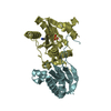
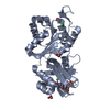
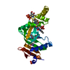
| Entry | Database: PDB / ID: 3m7f | ||||||
|---|---|---|---|---|---|---|---|
| Title | Crystal structure of the Nedd4 C2/Grb10 SH2 complex | ||||||
 Components Components |
| ||||||
 Keywords Keywords |  SIGNALING PROTEIN/LIGASE / Nedd4 / C2 domain / Grb10 / SH2 domain / Phosphoprotein / Ligase / Ubl conjugation pathway / SIGNALING PROTEIN-LIGASE complex SIGNALING PROTEIN/LIGASE / Nedd4 / C2 domain / Grb10 / SH2 domain / Phosphoprotein / Ligase / Ubl conjugation pathway / SIGNALING PROTEIN-LIGASE complex | ||||||
| Function / homology |  Function and homology information Function and homology informationRegulation of PTEN localization / Downregulation of ERBB4 signaling / IRS activation / Insulin receptor signalling cascade / Signal attenuation / ISG15 antiviral mechanism / Signaling by SCF-KIT / Regulation of PTEN stability and activity / negative regulation of sodium ion transport / endocardial cushion development ...Regulation of PTEN localization / Downregulation of ERBB4 signaling / IRS activation / Insulin receptor signalling cascade / Signal attenuation / ISG15 antiviral mechanism / Signaling by SCF-KIT / Regulation of PTEN stability and activity / negative regulation of sodium ion transport / endocardial cushion development / regulation of protein catabolic process at postsynapse, modulating synaptic transmission / : / negative regulation of sodium ion transmembrane transporter activity / negative regulation of glycogen biosynthetic process / phosphothreonine residue binding / receptor catabolic process / protein targeting to lysosome / RET signaling / Antigen processing: Ubiquitination & Proteasome degradation / negative regulation of phosphorylation / negative regulation of glucose import / HECT-type E3 ubiquitin transferase / sodium channel inhibitor activity / regulation of postsynaptic neurotransmitter receptor internalization / positive regulation of vascular endothelial growth factor receptor signaling pathway / proline-rich region binding / blood vessel morphogenesis / postsynaptic cytosol / neuromuscular junction development / regulation of dendrite morphogenesis / regulation of synapse organization / negative regulation of Wnt signaling pathway / negative regulation of vascular endothelial growth factor receptor signaling pathway / outflow tract morphogenesis / protein monoubiquitination / protein K63-linked ubiquitination / microvillus / phosphoserine residue binding / ubiquitin ligase complex / positive regulation of phosphorylation / ionotropic glutamate receptor binding / T cell activation / negative regulation of insulin receptor signaling pathway / phosphotyrosine residue binding / insulin-like growth factor receptor signaling pathway / response to insulin / insulin receptor binding / receptor internalization / protein polyubiquitination / positive regulation of protein catabolic process / ubiquitin-protein transferase activity / neuron projection development / ubiquitin protein ligase activity / signaling receptor complex adaptor activity / insulin receptor signaling pathway / protein-macromolecule adaptor activity / positive regulation of cold-induced thermogenesis / ubiquitin-dependent protein catabolic process / proteasome-mediated ubiquitin-dependent protein catabolic process / adaptive immune response / dendritic spine / protein ubiquitination / membrane raft / glutamatergic synapse / negative regulation of transcription by RNA polymerase II / signal transduction / protein-containing complex / membrane / identical protein binding / plasma membrane / cytosol / cytoplasmSimilarity search - Function | ||||||
| Biological species |  Mus musculus (house mouse) Mus musculus (house mouse) | ||||||
| Method | X-RAY DIFFRACTION / SYNCHROTRON / MOLECULAR REPLACEMENT / Resolution: 2 Å | ||||||
 Authors Authors | Huang, Q. / Szebenyi, M. | ||||||
 Citation Citation | Journal: J.Biol.Chem. / Year: 2010 Title: Structural Basis for the Interaction between the Growth Factor-binding Protein GRB10 and the E3 Ubiquitin Ligase NEDD4. Authors: Huang, Q. / Szebenyi, D.M. | ||||||
| History |
|
- Structure visualization
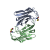
| Structure viewer | Molecule: MolmilJmol/JSmol |
|---|
- Downloads & links
-Download
| PDBx/mmCIF format | 3m7f.cif.gz | 116.2 KB | Display | PDBx/mmCIF format |
|---|---|---|---|---|
| PDB format | pdb3m7f.ent.gz | 89 KB | Display | PDB format |
| PDBx/mmJSON format | 3m7f.json.gz | Tree view | PDBx/mmJSON format | |
| Others |  Other downloads Other downloads |
-Validation report
| Arichive directory | https://data.pdbj.org/pub/pdb/validation_reports/m7/3m7fftp://data.pdbj.org/pub/pdb/validation_reports/m7/3m7f | HTTPS FTP |
|---|
-Related structure data


-Links
 PDBj
PDBj


- Assembly
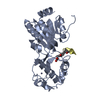
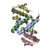
| Deposited unit | 
| ||||||||
|---|---|---|---|---|---|---|---|---|---|
| 1 |
| ||||||||
| Unit cell |
|
-Components
| #1: Protein | Mass: 12644.569 Da / Num. of mol.: 1 / Fragment: UNP residues 514-621, SH2 domain Source method: isolated from a genetically manipulated source Source: (gene. exp.) Mus musculus (house mouse) / Gene: Grb10, Kiaa0093, Meg1, Nedd-4, Nedd4, Nedd4a / Production host:  Escherichia coli (E. coli) / Strain (production host): BL21(DE3) Escherichia coli (E. coli) / Strain (production host): BL21(DE3)References: UniProt: Q60760, Ligases; Forming carbon-nitrogen bonds; Acid-amino-acid ligases (peptide synthases) |
|---|---|
| #2: Protein | Mass: 20339.244 Da / Num. of mol.: 1 / Fragment: UNP residues 71-246, C2 domain Source method: isolated from a genetically manipulated source Source: (gene. exp.) Mus musculus (house mouse) / Gene: Grb10, Kiaa0093, Meg1, Nedd-4, Nedd4, Nedd4a / Production host: Escherichia coli (E. coli) / Strain (production host): BL21(DE3)References: UniProt: P46935, Ligases; Forming carbon-nitrogen bonds; Acid-amino-acid ligases (peptide synthases) |
| #3: Water | ChemComp-HOH / Water Mass: 18.015 Da / Num. of mol.: 141 / Source method: isolated from a natural source / Formula: H2O Mass: 18.015 Da / Num. of mol.: 141 / Source method: isolated from a natural source / Formula: H2O |
| Sequence details | THIS RESIDUE AT POSITION 583 WAS IDENTIFIED |
-Experimental details
-Experiment
| Experiment | Method: X-RAY DIFFRACTION / Number of used crystals: 1 |
|---|
- Sample preparation
| Crystal | Density Matthews: 2.39 Å3/Da / Density % sol: 48.53 % |
|---|---|
| Crystal grow | Temperature: 295 K / Method: evaporation / pH: 6.5 Details: crystallization in 35% MPD, pH 6.5, EVAPORATION, temperature 295K |
-Data collection
| Diffraction | Mean temperature: 100 K |
|---|---|
| Diffraction source | Source: SYNCHROTRON / Site: CHESS  / Beamline: A1 / Wavelength: 0.978 Å / Beamline: A1 / Wavelength: 0.978 Å |
| Detector | Type: ADSC QUANTUM 210 / Detector: CCD / Date: Oct 28, 2009 / Details: mirrors |
| Radiation | Monochromator: mirrors / Protocol: SINGLE WAVELENGTH / Monochromatic (M) / Laue (L): M / Scattering type: x-ray |
| Radiation wavelength | Wavelength: 0.978 Å / Relative weight: 1 |
| Reflection | Resolution: 2→50 Å / Num. obs: 21866 / Redundancy: 6.5 % / Rmerge(I) obs: 0.052 |
- Processing
| Software |
| |||||||||||||||||||||||||||||||||||||||||||||||||||||||||||||||
|---|---|---|---|---|---|---|---|---|---|---|---|---|---|---|---|---|---|---|---|---|---|---|---|---|---|---|---|---|---|---|---|---|---|---|---|---|---|---|---|---|---|---|---|---|---|---|---|---|---|---|---|---|---|---|---|---|---|---|---|---|---|---|---|---|
| Refinement | Method to determine structure: MOLECULAR REPLACEMENT Starting model: PDB ENTRIES 1NRV AND 3B7Y Resolution: 2→43.123 Å / SU ML: 0.25 / σ(F): 0.06 / Stereochemistry target values: ML
| |||||||||||||||||||||||||||||||||||||||||||||||||||||||||||||||
| Solvent computation | Shrinkage radii: 0.9 Å / VDW probe radii: 1.11 Å / Solvent model: FLAT BULK SOLVENT MODEL / Bsol: 53.194 Å2 / ksol: 0.359 e/Å3 | |||||||||||||||||||||||||||||||||||||||||||||||||||||||||||||||
| Refine analyze | Luzzati sigma a obs: 0.25 Å | |||||||||||||||||||||||||||||||||||||||||||||||||||||||||||||||
| Refinement step | Cycle: LAST / Resolution: 2→43.123 Å
| |||||||||||||||||||||||||||||||||||||||||||||||||||||||||||||||
| Refine LS restraints |
| |||||||||||||||||||||||||||||||||||||||||||||||||||||||||||||||
| LS refinement shell |
| |||||||||||||||||||||||||||||||||||||||||||||||||||||||||||||||
| Refinement TLS params. | Method: refined / Origin x: 6.5699 Å / Origin y: 5.1861 Å / Origin z: 6.4036 Å
| |||||||||||||||||||||||||||||||||||||||||||||||||||||||||||||||
| Refinement TLS group | Selection details: all |
