 Movie
Movie Controller

[English] 日本語
 Yorodumi
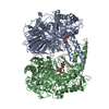
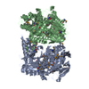
- PDB-3m46: The crystal structure of the D73A mutant of glycoside HYDROLASE (... -
+ Open data

- Basic information
| Entry | Database: PDB / ID: 3m46 | ||||||
|---|---|---|---|---|---|---|---|
| Title | The crystal structure of the D73A mutant of glycoside HYDROLASE (FAMILY 31) from Ruminococcus obeum ATCC 29174 | ||||||
 Components Components | Uncharacterized protein | ||||||
 Keywords Keywords |  HYDROLASE / Glycoside hydrolase (FAMILY 31) / structural genomics / PSI-2 / protein structure initiative / MCSG / Midwest Center for Structural Genomics HYDROLASE / Glycoside hydrolase (FAMILY 31) / structural genomics / PSI-2 / protein structure initiative / MCSG / Midwest Center for Structural Genomics | ||||||
| Function / homology |  Function and homology informationHydrolases; Glycosylases; Glycosidases, i.e. enzymes that hydrolyse O- and S-glycosyl compounds / hydrolase activity, hydrolyzing O-glycosyl compounds / carbohydrate binding / carbohydrate metabolic process Function and homology informationHydrolases; Glycosylases; Glycosidases, i.e. enzymes that hydrolyse O- and S-glycosyl compounds / hydrolase activity, hydrolyzing O-glycosyl compounds / carbohydrate binding / carbohydrate metabolic processSimilarity search - Function | ||||||
| Biological species |  Ruminococcus obeum (bacteria) Ruminococcus obeum (bacteria) | ||||||
| Method | X-RAY DIFFRACTION / SYNCHROTRON / MOLECULAR REPLACEMENT / Resolution: 2.66 Å | ||||||
 Authors Authors | Tan, K. / Tesar, C. / Freeman, L. / Babnigg, G. / Joachimiak, A. / Midwest Center for Structural Genomics (MCSG) | ||||||
 Citation Citation | Journal: Faseb J. / Year: 2010 Title: Novel alpha-glucosidase from human gut microbiome: substrate specificities and their switch Authors: Tan, K. / Tesar, C. / Wilton, R. / Keigher, L. / Babnigg, G. / Joachimiak, A. | ||||||
| History |
|
- Structure visualization
| Structure viewer | Molecule: MolmilJmol/JSmol |
|---|
- Downloads & links
-Download
| PDBx/mmCIF format | 3m46.cif.gz | 268.7 KB | Display | PDBx/mmCIF format |
|---|---|---|---|---|
| PDB format | pdb3m46.ent.gz | 216.1 KB | Display | PDB format |
| PDBx/mmJSON format | 3m46.json.gz | Tree view | PDBx/mmJSON format | |
| Others |  Other downloads Other downloads |
-Validation report
| Arichive directory | https://data.pdbj.org/pub/pdb/validation_reports/m4/3m46ftp://data.pdbj.org/pub/pdb/validation_reports/m4/3m46 | HTTPS FTP |
|---|
-Related structure data
| Related structure data | 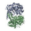 3m6dC  3mkkC  3n04C  3ffj C: citing same article ( S: Starting model for refinement |
|---|---|
| Similar structure data | |
| Other databases |


-Links
 PDBj
PDBj


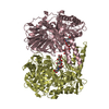
- Assembly
| Deposited unit | 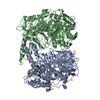
| ||||||||
|---|---|---|---|---|---|---|---|---|---|
| 1 |
| ||||||||
| Unit cell |
|
-Components
| #1: Protein | Mass: 77405.453 Da / Num. of mol.: 2 / Mutation: D73A Source method: isolated from a genetically manipulated source Source: (gene. exp.) Ruminococcus obeum (bacteria) / Strain: ATCC 29174 / Gene: RUMOBE_03919 / Plasmid: pMCSG19 / Production host: Escherichia coli (E. coli) / Strain (production host): pPK1037 / References: UniProt: A5ZY13#2: Chemical | ChemComp-GOL / | Glycerol  Mass: 92.094 Da / Num. of mol.: 1 / Source method: obtained synthetically / Formula: C3H8O3 Mass: 92.094 Da / Num. of mol.: 1 / Source method: obtained synthetically / Formula: C3H8O3#3: Water | ChemComp-HOH / | Water Mass: 18.015 Da / Num. of mol.: 52 / Source method: isolated from a natural source / Formula: H2O Mass: 18.015 Da / Num. of mol.: 52 / Source method: isolated from a natural source / Formula: H2O |
|---|
-Experimental details
-Experiment
| Experiment | Method: X-RAY DIFFRACTION / Number of used crystals: 1 |
|---|
- Sample preparation
| Crystal | Density Matthews: 2.1 Å3/Da / Density % sol: 41.56 % |
|---|---|
| Crystal grow | Temperature: 289 K / Method: vapor diffusion, sitting drop / pH: 5.5 Details: 0.1M BIS-TRIS, 25% PEG3350, 5mM maltose, pH 5.5, VAPOR DIFFUSION, SITTING DROP, temperature 289K |
-Data collection
| Diffraction | Mean temperature: 100 K |
|---|---|
| Diffraction source | Source: SYNCHROTRON / Site: APS  / Beamline: 19-ID / Wavelength: 0.97951 Å / Beamline: 19-ID / Wavelength: 0.97951 Å |
| Detector | Type: ADSC QUANTUM 315r / Detector: CCD / Date: Feb 24, 2010 / Details: Mirror |
| Radiation | Monochromator: Si 111 crystal / Protocol: SINGLE WAVELENGTH / Monochromatic (M) / Laue (L): M / Scattering type: x-ray |
| Radiation wavelength | Wavelength: 0.97951 Å / Relative weight: 1 |
| Reflection | Resolution: 2.65→83 Å / Num. all: 36534 / Num. obs: 36534 / % possible obs: 99.5 % / Observed criterion σ(F): 0 / Observed criterion σ(I): 0 / Redundancy: 3.5 % / Rmerge(I) obs: 0.143 / Net I/σ(I): 8.8 |
| Reflection shell | Resolution: 2.65→2.7 Å / Redundancy: 3.4 % / Rmerge(I) obs: 0.742 / Mean I/σ(I) obs: 1.45 / Num. unique all: 2001 / % possible all: 99.2 |
- Processing
| Software |
| ||||||||||||||||||||||||||||||||||||||||||||||||||||||||||||||||||||||||||||||||||||||||||||||||||||||||||||||||||||||||||||||||||||||||||||||||||||||||||||||||||||||||||
|---|---|---|---|---|---|---|---|---|---|---|---|---|---|---|---|---|---|---|---|---|---|---|---|---|---|---|---|---|---|---|---|---|---|---|---|---|---|---|---|---|---|---|---|---|---|---|---|---|---|---|---|---|---|---|---|---|---|---|---|---|---|---|---|---|---|---|---|---|---|---|---|---|---|---|---|---|---|---|---|---|---|---|---|---|---|---|---|---|---|---|---|---|---|---|---|---|---|---|---|---|---|---|---|---|---|---|---|---|---|---|---|---|---|---|---|---|---|---|---|---|---|---|---|---|---|---|---|---|---|---|---|---|---|---|---|---|---|---|---|---|---|---|---|---|---|---|---|---|---|---|---|---|---|---|---|---|---|---|---|---|---|---|---|---|---|---|---|---|---|---|---|
| Refinement | Method to determine structure: MOLECULAR REPLACEMENT Starting model: PDB entry 3FFJ 3ffj Resolution: 2.66→82.89 Å / Cor.coef. Fo:Fc: 0.93 / Cor.coef. Fo:Fc free: 0.889 / SU B: 30.235 / SU ML: 0.281 / TLS residual ADP flag: LIKELY RESIDUAL / Cross valid method: THROUGHOUT / σ(F): 0 / ESU R Free: 0.389 / Stereochemistry target values: MAXIMUM LIKELIHOOD / Details: HYDROGENS HAVE BEEN ADDED IN THE RIDING POSITIONS
| ||||||||||||||||||||||||||||||||||||||||||||||||||||||||||||||||||||||||||||||||||||||||||||||||||||||||||||||||||||||||||||||||||||||||||||||||||||||||||||||||||||||||||
| Solvent computation | Ion probe radii: 0.8 Å / Shrinkage radii: 0.8 Å / VDW probe radii: 1.4 Å / Solvent model: MASK | ||||||||||||||||||||||||||||||||||||||||||||||||||||||||||||||||||||||||||||||||||||||||||||||||||||||||||||||||||||||||||||||||||||||||||||||||||||||||||||||||||||||||||
| Displacement parameters | Biso mean: 37.13 Å2
| ||||||||||||||||||||||||||||||||||||||||||||||||||||||||||||||||||||||||||||||||||||||||||||||||||||||||||||||||||||||||||||||||||||||||||||||||||||||||||||||||||||||||||
| Refinement step | Cycle: LAST / Resolution: 2.66→82.89 Å
| ||||||||||||||||||||||||||||||||||||||||||||||||||||||||||||||||||||||||||||||||||||||||||||||||||||||||||||||||||||||||||||||||||||||||||||||||||||||||||||||||||||||||||
| Refine LS restraints |
| ||||||||||||||||||||||||||||||||||||||||||||||||||||||||||||||||||||||||||||||||||||||||||||||||||||||||||||||||||||||||||||||||||||||||||||||||||||||||||||||||||||||||||
| LS refinement shell | Resolution: 2.659→2.728 Å / Total num. of bins used: 20
| ||||||||||||||||||||||||||||||||||||||||||||||||||||||||||||||||||||||||||||||||||||||||||||||||||||||||||||||||||||||||||||||||||||||||||||||||||||||||||||||||||||||||||
| Refinement TLS params. | Method: refined / Refine-ID: X-RAY DIFFRACTION
| ||||||||||||||||||||||||||||||||||||||||||||||||||||||||||||||||||||||||||||||||||||||||||||||||||||||||||||||||||||||||||||||||||||||||||||||||||||||||||||||||||||||||||
| Refinement TLS group |
|
