 Movie
Movie Controller

[English] 日本語
 Yorodumi
- PDB-3lyu: Crystal Structure of the C-terminal domain (residues 83-215) of P... -
+ Open data

- Basic information
| Entry | Database: PDB / ID: 3lyu | ||||||
|---|---|---|---|---|---|---|---|
| Title | Crystal Structure of the C-terminal domain (residues 83-215) of PF1911 hydrogenase from Pyrococcus furiosus, Northeast Structural Genomics Consortium Target PfR246A | ||||||
 Components Components | Putative hydrogenase | ||||||
 Keywords Keywords | OXIDOREDUCTASE / The C-terminal has an alpha-beta fold / Structural Genomics / PSI-2 / Protein Structure Initiative / Northeast Structural Genomics Consortium / NESG | ||||||
| Function / homology |  Function and homology information Function and homology informationpyrimidine nucleotide biosynthetic process / 2 iron, 2 sulfur cluster binding / flavin adenine dinucleotide binding / oxidoreductase activity / metal ion bindingSimilarity search - Function | ||||||
| Biological species |   Pyrococcus furiosus (archaea) Pyrococcus furiosus (archaea) | ||||||
| Method | X-RAY DIFFRACTION / SYNCHROTRON / SAD / Resolution: 2.3 Å | ||||||
 Authors Authors | Forouhar, F. / Abashidze, M. / Seetharaman, J. / Sahdev, S. / Xiao, R. / Foote, E.L. / Ciccosanti, C. / Belote, R.L. / Everett, J.K. / Nair, R. ...Forouhar, F. / Abashidze, M. / Seetharaman, J. / Sahdev, S. / Xiao, R. / Foote, E.L. / Ciccosanti, C. / Belote, R.L. / Everett, J.K. / Nair, R. / Acton, T.B. / Rost, B. / Montelione, G.T. / Tong, L. / Hunt, J.F. / Northeast Structural Genomics Consortium (NESG) | ||||||
 Citation Citation | Journal: To be Published Title: Northeast Structural Genomics Consortium Target PfR246A Authors: Forouhar, F. / Abashidze, M. / Seetharaman, J. / Sahdev, S. / Xiao, R. / Foote, E.L. / Ciccosanti, C. / Belote, R.L. / Everett, J.K. / Nair, R. / Acton, T.B. / Rost, B. / Montelione, G.T. / Tong, L. / Hunt, J.F. | ||||||
| History |
|
- Structure visualization
| Structure viewer | Molecule: MolmilJmol/JSmol |
|---|
- Downloads & links
-Download
| PDBx/mmCIF format | 3lyu.cif.gz | 158.4 KB | Display | PDBx/mmCIF format |
|---|---|---|---|---|
| PDB format | pdb3lyu.ent.gz | 133.4 KB | Display | PDB format |
| PDBx/mmJSON format | 3lyu.json.gz | Tree view | PDBx/mmJSON format | |
| Others |  Other downloads Other downloads |
-Validation report
| Arichive directory | https://data.pdbj.org/pub/pdb/validation_reports/ly/3lyuftp://data.pdbj.org/pub/pdb/validation_reports/ly/3lyu | HTTPS FTP |
|---|
-Related structure data


-Links
 PDBj
PDBj- Assembly
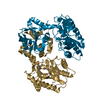
| Deposited unit | 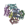
| ||||||||
|---|---|---|---|---|---|---|---|---|---|
| 1 |
| ||||||||
| 2 | 
| ||||||||
| 3 | 
| ||||||||
| 4 | 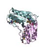
| ||||||||
| Unit cell |
| ||||||||
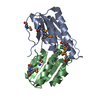
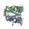
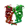
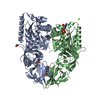
| Details | According to the static light scattering data, the C-terminal hydrogenase is dimer in solution, whereas in the crystal forms a hexameric ring. |
-Components
| #1: Protein | Mass: 16413.072 Da / Num. of mol.: 6 / Fragment: sequence database residues 83-215 Source method: isolated from a genetically manipulated source Source: (gene. exp.) Pyrococcus furiosus (archaea) / Strain: DSM 3638 / Gene: PF1911 / Production host:  Escherichia coli (E. coli) / Strain (production host): BL21(DE3)+ Magic / References: UniProt: Q8TZS3 Escherichia coli (E. coli) / Strain (production host): BL21(DE3)+ Magic / References: UniProt: Q8TZS3#2: Water | ChemComp-HOH / | Water Mass: 18.015 Da / Num. of mol.: 69 / Source method: isolated from a natural source / Formula: H2O Mass: 18.015 Da / Num. of mol.: 69 / Source method: isolated from a natural source / Formula: H2O |
|---|
-Experimental details
-Experiment
| Experiment | Method: X-RAY DIFFRACTION / Number of used crystals: 1 |
|---|
- Sample preparation
| Crystal | Density Matthews: 2.08 Å3/Da / Density % sol: 40.79 % |
|---|---|
| Crystal grow | Temperature: 291 K / Method: vapor diffusion, hanging drop / pH: 6.5 Details: Protein solution: 100mM NaCl, 5mM DTT, 0.02% NaN3, 10mM Tris-HCl (pH 7.5), Reservoir solution: 0.1M MES (pH 6.5), 18% PEG3350, and 0.2M calcium chloride, VAPOR DIFFUSION, HANGING DROP, temperature 291K |
-Data collection
| Diffraction | Mean temperature: 100 K | |||||||||||||||
|---|---|---|---|---|---|---|---|---|---|---|---|---|---|---|---|---|
| Diffraction source | Source: SYNCHROTRON / Site: NSLS  / Beamline: X4C / Wavelength: 0.97885 Å / Beamline: X4C / Wavelength: 0.97885 Å | |||||||||||||||
| Detector | Type: MAR CCD 165 mm / Detector: CCD / Date: Apr 8, 2009 / Details: mirrors | |||||||||||||||
| Radiation | Monochromator: Si 111 CHANNEL / Protocol: SINGLE WAVELENGTH / Monochromatic (M) / Laue (L): M / Scattering type: x-ray | |||||||||||||||
| Radiation wavelength | Wavelength: 0.97885 Å / Relative weight: 1 | |||||||||||||||
| Reflection twin |
| |||||||||||||||
| Reflection | Resolution: 2.3→30 Å / Num. all: 70503 / Num. obs: 70362 / % possible obs: 99.8 % / Observed criterion σ(F): 0 / Observed criterion σ(I): 0 / Redundancy: 5.2 % / Biso Wilson estimate: 24.5 Å2 / Rmerge(I) obs: 0.092 / Rsym value: 0.088 / Net I/σ(I): 20.8 | |||||||||||||||
| Reflection shell | Resolution: 2.3→2.38 Å / Redundancy: 5.1 % / Rmerge(I) obs: 0.338 / Mean I/σ(I) obs: 3.4 / Num. unique all: 6941 / Rsym value: 0.368 / % possible all: 99.7 |
- Processing
| Software |
| ||||||||||||||||||||||||||||||||
|---|---|---|---|---|---|---|---|---|---|---|---|---|---|---|---|---|---|---|---|---|---|---|---|---|---|---|---|---|---|---|---|---|---|
| Refinement | Method to determine structure: SAD / Resolution: 2.3→19.59 Å / Rfactor Rfree error: 0.005 / Data cutoff high absF: 396034.719 / Data cutoff low absF: 0 / Isotropic thermal model: RESTRAINED / Cross valid method: THROUGHOUT / σ(F): 2 / σ(I): 2 / Stereochemistry target values: Engh & Huber Details: TWIN DETAILS NUMBER OF TWIN DOMAINS : 2 TWIN DOMAIN : 1 TWIN OPERATOR : H, K, L TWIN FRACTION : 0.497 TWIN DOMAIN : 2 TWIN OPERATOR : K, H, -L TWIN FRACTION : 0.503
| ||||||||||||||||||||||||||||||||
| Solvent computation | Solvent model: FLAT MODEL / Bsol: 41.276 Å2 / ksol: 0.4 e/Å3 | ||||||||||||||||||||||||||||||||
| Displacement parameters | Biso mean: 46.2 Å2
| ||||||||||||||||||||||||||||||||
| Refine analyze |
| ||||||||||||||||||||||||||||||||
| Refinement step | Cycle: LAST / Resolution: 2.3→19.59 Å
| ||||||||||||||||||||||||||||||||
| Refine LS restraints |
| ||||||||||||||||||||||||||||||||
| LS refinement shell | Resolution: 2.3→2.38 Å / Rfactor Rfree error: 0.017 / Total num. of bins used: 10
|
