 Movie
Movie Controller

[English] 日本語
 Yorodumi
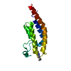
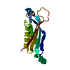
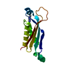
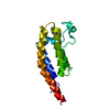
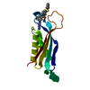
- PDB-3lq9: Crystal structure of human REDD1, a hypoxia-induced regulator of mTOR -
+ Open data

- Basic information
| Entry | Database: PDB / ID: 3lq9 | ||||||
|---|---|---|---|---|---|---|---|
| Title | Crystal structure of human REDD1, a hypoxia-induced regulator of mTOR | ||||||
 Components Components | DNA-damage-inducible transcript 4 protein | ||||||
 Keywords Keywords |  SIGNALING PROTEIN / REDD1 DDIT4 mTOR / hypoxia / cancer SIGNALING PROTEIN / REDD1 DDIT4 mTOR / hypoxia / cancer | ||||||
| Function / homology |  Function and homology information Function and homology informationprotein-containing complex disassembly / negative regulation of glycolytic process / negative regulation of TOR signaling / neurotrophin TRK receptor signaling pathway / intrinsic apoptotic signaling pathway in response to DNA damage by p53 class mediator / 14-3-3 protein binding / reactive oxygen species metabolic process / cellular response to dexamethasone stimulus / TP53 Regulates Metabolic Genes / neuron migration ...protein-containing complex disassembly / negative regulation of glycolytic process / negative regulation of TOR signaling / neurotrophin TRK receptor signaling pathway / intrinsic apoptotic signaling pathway in response to DNA damage by p53 class mediator / 14-3-3 protein binding / reactive oxygen species metabolic process / cellular response to dexamethasone stimulus / TP53 Regulates Metabolic Genes / neuron migration / brain development / neuron differentiation / defense response to virus / response to hypoxia / intracellular signal transduction / apoptotic process / mitochondrion / cytosol / cytoplasmSimilarity search - Function | ||||||
| Biological species |  Homo sapiens (human) Homo sapiens (human) | ||||||
| Method | X-RAY DIFFRACTION / SYNCHROTRON / SAD / Resolution: 2 Å | ||||||
 Authors Authors | Vega-Rubin-de-Celis, S. / Abdallah, Z. / Brugarolas, J. / Zhang, X. | ||||||
 Citation Citation | Journal: Biochemistry / Year: 2010 Title: Structural analysis and functional implications of the negative mTORC1 regulator REDD1. Authors: Vega-Rubin-de-Celis, S. / Abdallah, Z. / Kinch, L. / Grishin, N.V. / Brugarolas, J. / Zhang, X. | ||||||
| History |
|
- Structure visualization
| Structure viewer | Molecule: MolmilJmol/JSmol |
|---|
- Downloads & links
-Download
| PDBx/mmCIF format | 3lq9.cif.gz | 61.8 KB | Display | PDBx/mmCIF format |
|---|---|---|---|---|
| PDB format | pdb3lq9.ent.gz | 48.3 KB | Display | PDB format |
| PDBx/mmJSON format | 3lq9.json.gz | Tree view | PDBx/mmJSON format | |
| Others |  Other downloads Other downloads |
-Validation report
| Arichive directory | https://data.pdbj.org/pub/pdb/validation_reports/lq/3lq9ftp://data.pdbj.org/pub/pdb/validation_reports/lq/3lq9 | HTTPS FTP |
|---|
-Related structure data
| Similar structure data |
|---|


-Links
 PDBj
PDBj
- Assembly
| Deposited unit | 
| ||||||||
|---|---|---|---|---|---|---|---|---|---|
| 1 | 
| ||||||||
| 2 | 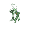
| ||||||||
| Unit cell |
|
-Components
| #1: Protein | Mass: 14830.898 Da / Num. of mol.: 2 / Fragment: C-terminal functional domain / Mutation: deletion of residues 200-204 Source method: isolated from a genetically manipulated source Source: (gene. exp.) Homo sapiens (human) / Gene: DDIT4, REDD1, RTP801 / Plasmid: modified pET28 with a His6-Sumo tag / Production host:  Escherichia coli (E. coli) / Strain (production host): BL21(DE3) / References: UniProt: Q9NX09 Escherichia coli (E. coli) / Strain (production host): BL21(DE3) / References: UniProt: Q9NX09#2: Water | ChemComp-HOH / | Water Mass: 18.015 Da / Num. of mol.: 203 / Source method: isolated from a natural source / Formula: H2O Mass: 18.015 Da / Num. of mol.: 203 / Source method: isolated from a natural source / Formula: H2O |
|---|
-Experimental details
-Experiment
| Experiment | Method: X-RAY DIFFRACTION / Number of used crystals: 2 |
|---|
- Sample preparation
| Crystal | Density Matthews: 1.92 Å3/Da / Density % sol: 35.99 % |
|---|---|
| Crystal grow | Temperature: 277 K / Method: vapor diffusion, hanging drop Details: 0.1 M NaF, 20-26% PEG3350, 0.05 mM C12E9, vapor diffusion, hanging drop, temperature 277K |
-Data collection
| Diffraction |
| |||||||||||||||||||||||||||||||||||||||||||||||||||||||||||||||||||||||||||||
|---|---|---|---|---|---|---|---|---|---|---|---|---|---|---|---|---|---|---|---|---|---|---|---|---|---|---|---|---|---|---|---|---|---|---|---|---|---|---|---|---|---|---|---|---|---|---|---|---|---|---|---|---|---|---|---|---|---|---|---|---|---|---|---|---|---|---|---|---|---|---|---|---|---|---|---|---|---|---|
| Diffraction source |
| |||||||||||||||||||||||||||||||||||||||||||||||||||||||||||||||||||||||||||||
| Detector |
| |||||||||||||||||||||||||||||||||||||||||||||||||||||||||||||||||||||||||||||
| Radiation |
| |||||||||||||||||||||||||||||||||||||||||||||||||||||||||||||||||||||||||||||
| Radiation wavelength |
| |||||||||||||||||||||||||||||||||||||||||||||||||||||||||||||||||||||||||||||
| Reflection | Redundancy: 4.7 % / Av σ(I) over netI: 25.34 / Number: 55284 / Rmerge(I) obs: 0.079 / Χ2: 1.93 / D res high: 2.2 Å / D res low: 50 Å / Num. obs: 11682 / % possible obs: 96 | |||||||||||||||||||||||||||||||||||||||||||||||||||||||||||||||||||||||||||||
| Diffraction reflection shell |
| |||||||||||||||||||||||||||||||||||||||||||||||||||||||||||||||||||||||||||||
| Reflection | Resolution: 2→50 Å / Num. obs: 14319 / % possible obs: 95.9 % / Redundancy: 4 % / Rmerge(I) obs: 0.068 / Χ2: 1.859 / Net I/σ(I): 15.9 | |||||||||||||||||||||||||||||||||||||||||||||||||||||||||||||||||||||||||||||
| Reflection shell |
|

- Processing
| Software |
| ||||||||||||||||||||||||||||||||||||||||||
|---|---|---|---|---|---|---|---|---|---|---|---|---|---|---|---|---|---|---|---|---|---|---|---|---|---|---|---|---|---|---|---|---|---|---|---|---|---|---|---|---|---|---|---|
| Refinement | Method to determine structure: SAD / Resolution: 2→27.096 Å / Occupancy max: 1 / Occupancy min: 1 / FOM work R set: 0.851 / SU ML: 0.28 / σ(F): 1 / Stereochemistry target values: MLHL
| ||||||||||||||||||||||||||||||||||||||||||
| Solvent computation | Shrinkage radii: 0.9 Å / VDW probe radii: 1.11 Å / Solvent model: FLAT BULK SOLVENT MODEL / Bsol: 48.587 Å2 / ksol: 0.365 e/Å3 | ||||||||||||||||||||||||||||||||||||||||||
| Displacement parameters | Biso max: 90.6 Å2 / Biso mean: 30.4 Å2 / Biso min: 11.37 Å2
| ||||||||||||||||||||||||||||||||||||||||||
| Refinement step | Cycle: LAST / Resolution: 2→27.096 Å
| ||||||||||||||||||||||||||||||||||||||||||
| Refine LS restraints |
| ||||||||||||||||||||||||||||||||||||||||||
| LS refinement shell | Refine-ID: X-RAY DIFFRACTION / Total num. of bins used: 5
|
