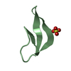
 Movie
Movie Controller


+ Open data

- Basic information
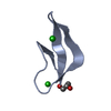
| Entry | Database: PDB / ID: 3lo9 | ||||||
|---|---|---|---|---|---|---|---|
| Title | Crystal structure of human alpha-defensin 1 (W26Ahp mutant) | ||||||
 Components Components | Neutrophil defensin 1 | ||||||
 Keywords Keywords |  ANTIMICROBIAL PROTEIN / ANTIMICROBIAL PEPTIDE / HUMAN ALPHA DEFENSIN 1 / HUMAN NEUTROPHIL PEPTIDE 1 / HNP1 / ANTIBIOTIC / ANTIMICROBIAL / Antiviral defense / Defensin / Disulfide bond / Fungicide / Phosphoprotein / Secreted ANTIMICROBIAL PROTEIN / ANTIMICROBIAL PEPTIDE / HUMAN ALPHA DEFENSIN 1 / HUMAN NEUTROPHIL PEPTIDE 1 / HNP1 / ANTIBIOTIC / ANTIMICROBIAL / Antiviral defense / Defensin / Disulfide bond / Fungicide / Phosphoprotein / Secreted | ||||||
| Function / homology |  Function and homology information Function and homology informationpore-forming activity / Defensins / disruption of plasma membrane integrity in another organism / killing by host of symbiont cells / T cell chemotaxis / Alpha-defensins / defense response to protozoan / intracellular estrogen receptor signaling pathway / defense response to fungus / innate immune response in mucosa ...pore-forming activity / Defensins / disruption of plasma membrane integrity in another organism / killing by host of symbiont cells / T cell chemotaxis / Alpha-defensins / defense response to protozoan / intracellular estrogen receptor signaling pathway / defense response to fungus / innate immune response in mucosa / Golgi lumen / chemotaxis / azurophil granule lumen / antimicrobial humoral immune response mediated by antimicrobial peptide / antibacterial humoral response / collagen-containing extracellular matrix / defense response to virus / killing of cells of another organism / cellular response to lipopolysaccharide / defense response to Gram-negative bacterium / defense response to Gram-positive bacterium / immune response / Neutrophil degranulation / extracellular space / extracellular exosome / extracellular regionSimilarity search - Function | ||||||
| Method | X-RAY DIFFRACTION / MOLECULAR REPLACEMENT / Resolution: 1.56 Å | ||||||
 Authors Authors | Pazgier, M. / Lu, W. | ||||||
 Citation Citation | Journal: J.Biol.Chem. / Year: 2010 Title: Trp-26 imparts functional versatility to human alpha-defensin HNP1. Authors: Wei, G. / Pazgier, M. / de Leeuw, E. / Rajabi, M. / Li, J. / Zou, G. / Jung, G. / Yuan, W. / Lu, W.Y. / Lehrer, R.I. / Lu, W. | ||||||
| History |
|
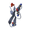
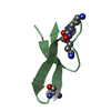
- Structure visualization
| Structure viewer | Molecule: MolmilJmol/JSmol |
|---|
- Downloads & links
-Download
| PDBx/mmCIF format | 3lo9.cif.gz | 24.9 KB | Display | PDBx/mmCIF format |
|---|---|---|---|---|
| PDB format | pdb3lo9.ent.gz | 16.1 KB | Display | PDB format |
| PDBx/mmJSON format | 3lo9.json.gz | Tree view | PDBx/mmJSON format | |
| Others |  Other downloads Other downloads |
-Validation report
| Arichive directory | https://data.pdbj.org/pub/pdb/validation_reports/lo/3lo9ftp://data.pdbj.org/pub/pdb/validation_reports/lo/3lo9 | HTTPS FTP |
|---|
-Related structure data
| Related structure data | 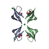 3h6cC 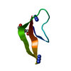 3lo1C 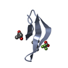 3lo2C  3lo4C 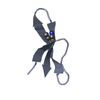 3lo6C 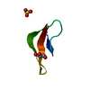 3loeC 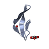 3lvxC  3gnyS S: Starting model for refinement C: citing same article ( |
|---|---|
| Similar structure data |


-Links
 PDBj
PDBj


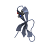
- Assembly
| Deposited unit | 
| ||||||||||||||||||
|---|---|---|---|---|---|---|---|---|---|---|---|---|---|---|---|---|---|---|---|
| 1 | 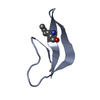
| ||||||||||||||||||
| 2 | 
| ||||||||||||||||||
| 3 |
| ||||||||||||||||||
| Unit cell |
| ||||||||||||||||||
| Noncrystallographic symmetry (NCS) | NCS domain:
NCS domain segments: Component-ID: 1 / Ens-ID: 1 / Beg auth comp-ID: ALA / Beg label comp-ID: ALA / End auth comp-ID: CYS / End label comp-ID: CYS / Refine code: 6 / Auth seq-ID: 1 - 30 / Label seq-ID: 1 - 30
| ||||||||||||||||||
| Details | biological unit is half of asymmetric unit. |
-Components
| #1: Protein/peptide | Mass: 3393.085 Da / Num. of mol.: 2 / Fragment: UNP residues 65-94 / Mutation: W26Ahp / Source method: obtained synthetically / Details: Protein naturally occurs in HUMAN / References: UniProt: P59665 #2: Water | ChemComp-HOH / | Water Mass: 18.015 Da / Num. of mol.: 62 / Source method: isolated from a natural source / Formula: H2O Mass: 18.015 Da / Num. of mol.: 62 / Source method: isolated from a natural source / Formula: H2O |
|---|
-Experimental details
-Experiment
| Experiment | Method: X-RAY DIFFRACTION / Number of used crystals: 1 |
|---|
- Sample preparation
| Crystal | Density Matthews: 2.07 Å3/Da / Density % sol: 40.71 % |
|---|---|
| Crystal grow | Temperature: 298 K / Method: vapor diffusion, hanging drop / pH: 6.5 Details: 0.1 M sodium cacodylate trihydrate pH 6.5; 0.2 M sodium citrate tribasic dehydrate; 30% isopropanol , VAPOR DIFFUSION, HANGING DROP, temperature 298K |
-Data collection
| Diffraction | Mean temperature: 100 K |
|---|---|
| Diffraction source | Source: ROTATING ANODE / Type: RIGAKU MICROMAX-007 / Wavelength: 1.54 Å |
| Detector | Type: RIGAKU RAXIS IV++ / Detector: IMAGE PLATE / Date: Oct 6, 2008 |
| Radiation | Protocol: SINGLE WAVELENGTH / Monochromatic (M) / Laue (L): M / Scattering type: x-ray |
| Radiation wavelength | Wavelength: 1.54 Å / Relative weight: 1 |
| Reflection | Resolution: 1.56→30.643 Å / Num. all: 8440 / Num. obs: 8232 / % possible obs: 98 % / Observed criterion σ(F): 1 / Observed criterion σ(I): 1 / Redundancy: 6.3 % / Rmerge(I) obs: 0.059 / Rsym value: 0.081 / Net I/σ(I): 20.7 |
| Reflection shell | Resolution: 1.56→1.62 Å / Redundancy: 6.2 % / Rmerge(I) obs: 0.132 / Mean I/σ(I) obs: 18.8 / Num. unique all: 737 / Rsym value: 0.141 / % possible all: 88.2 |
- Processing
| Software |
| |||||||||||||||||||||||||||||||||||||||||||||||||||||||||||||||||||||||||||
|---|---|---|---|---|---|---|---|---|---|---|---|---|---|---|---|---|---|---|---|---|---|---|---|---|---|---|---|---|---|---|---|---|---|---|---|---|---|---|---|---|---|---|---|---|---|---|---|---|---|---|---|---|---|---|---|---|---|---|---|---|---|---|---|---|---|---|---|---|---|---|---|---|---|---|---|---|
| Refinement | Method to determine structure: MOLECULAR REPLACEMENT Starting model: PDB ENTRY 3GNY Resolution: 1.56→15 Å / Cor.coef. Fo:Fc: 0.964 / Cor.coef. Fo:Fc free: 0.948 / Occupancy max: 1 / Occupancy min: 1 / SU B: 3.411 / SU ML: 0.055 / TLS residual ADP flag: LIKELY RESIDUAL / Cross valid method: THROUGHOUT / σ(F): 0 / ESU R: 0.092 / ESU R Free: 0.086 / Stereochemistry target values: MAXIMUM LIKELIHOOD / Details: U VALUES : RESIDUAL ONLY
| |||||||||||||||||||||||||||||||||||||||||||||||||||||||||||||||||||||||||||
| Solvent computation | Ion probe radii: 0.8 Å / Shrinkage radii: 0.8 Å / VDW probe radii: 1.2 Å / Solvent model: MASK | |||||||||||||||||||||||||||||||||||||||||||||||||||||||||||||||||||||||||||
| Displacement parameters | Biso max: 42.57 Å2 / Biso mean: 12.556 Å2 / Biso min: 4.6 Å2
| |||||||||||||||||||||||||||||||||||||||||||||||||||||||||||||||||||||||||||
| Refinement step | Cycle: LAST / Resolution: 1.56→15 Å
| |||||||||||||||||||||||||||||||||||||||||||||||||||||||||||||||||||||||||||
| Refine LS restraints |
| |||||||||||||||||||||||||||||||||||||||||||||||||||||||||||||||||||||||||||
| Refine LS restraints NCS | Dom-ID: 1 / Auth asym-ID: A / Ens-ID: 1 / Number: 233 / Refine-ID: X-RAY DIFFRACTION
| |||||||||||||||||||||||||||||||||||||||||||||||||||||||||||||||||||||||||||
| LS refinement shell | Resolution: 1.563→1.603 Å / Total num. of bins used: 20
| |||||||||||||||||||||||||||||||||||||||||||||||||||||||||||||||||||||||||||
| Refinement TLS params. | Method: refined / Refine-ID: X-RAY DIFFRACTION
| |||||||||||||||||||||||||||||||||||||||||||||||||||||||||||||||||||||||||||
| Refinement TLS group |
|
