 Movie
Movie Controller

[English] 日本語
 Yorodumi
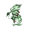
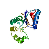
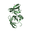
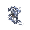
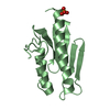
- PDB-3lax: The crystal structure of a domain of phenylacetate-coenzyme A lig... -
+ Open data

- Basic information
| Entry | Database: PDB / ID: 3lax | |||||||||
|---|---|---|---|---|---|---|---|---|---|---|
| Title | The crystal structure of a domain of phenylacetate-coenzyme A ligase from Bacteroides vulgatus ATCC 8482 | |||||||||
 Components Components | Phenylacetate-coenzyme A ligase | |||||||||
 Keywords Keywords |  LIGASE / structural genomics / PSI-2 / protein structure initiative / midwest center for structural genomics / MCSG LIGASE / structural genomics / PSI-2 / protein structure initiative / midwest center for structural genomics / MCSG | |||||||||
| Function / homology |  Function and homology informationphenylacetate-CoA ligase / phenylacetate-CoA ligase activity / phenylacetate catabolic process / nucleotide binding Function and homology informationphenylacetate-CoA ligase / phenylacetate-CoA ligase activity / phenylacetate catabolic process / nucleotide bindingSimilarity search - Function | |||||||||
| Biological species |  Bacteroides vulgatus (bacteria) Bacteroides vulgatus (bacteria) | |||||||||
| Method | X-RAY DIFFRACTION / SYNCHROTRON / SAD / Resolution: 1.428 Å | |||||||||
 Authors Authors | Tan, K. / Wu, R. / Cobb, G. / Joachimiak, A. / Midwest Center for Structural Genomics (MCSG) | |||||||||
 Citation Citation | Journal: To be Published Title: The crystal structure of a domain of phenylacetate-coenzyme A ligase from Bacteroides vulgatus ATCC 8482 Authors: Tan, K. / Wu, R. / Cobb, G. / Joachimiak, A. | |||||||||
| History |
|
- Structure visualization
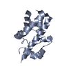
| Structure viewer | Molecule: MolmilJmol/JSmol |
|---|
- Downloads & links
-Download
| PDBx/mmCIF format | 3lax.cif.gz | 57.5 KB | Display | PDBx/mmCIF format |
|---|---|---|---|---|
| PDB format | pdb3lax.ent.gz | 46.4 KB | Display | PDB format |
| PDBx/mmJSON format | 3lax.json.gz | Tree view | PDBx/mmJSON format | |
| Others |  Other downloads Other downloads |
-Validation report
| Arichive directory | https://data.pdbj.org/pub/pdb/validation_reports/la/3laxftp://data.pdbj.org/pub/pdb/validation_reports/la/3lax | HTTPS FTP |
|---|
-Related structure data
| Similar structure data | |
|---|---|
| Other databases |


-Links
 PDBj
PDBj
- Assembly
| Deposited unit | 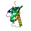
| ||||||||
|---|---|---|---|---|---|---|---|---|---|
| 1 |
| ||||||||
| Unit cell |
| ||||||||
| Details | Experimentally unknown. The domain is likely a monomer. |
-Components
| #1: Protein | Mass: 12473.078 Da / Num. of mol.: 1 / Fragment: residues 327-432 Source method: isolated from a genetically manipulated source Source: (gene. exp.) Bacteroides vulgatus (bacteria) / Strain: ATCC 8482 / Gene: Bacteroides vulgatus, BVU_1668 / Plasmid: pMCSG19 / Production host: Escherichia coli (E. coli) / Strain (production host): pPK1037 / References: UniProt: A6L0Y5 |
|---|---|
| #2: Water | ChemComp-HOH / Water Mass: 18.015 Da / Num. of mol.: 90 / Source method: isolated from a natural source / Formula: H2O Mass: 18.015 Da / Num. of mol.: 90 / Source method: isolated from a natural source / Formula: H2O |
-Experimental details
-Experiment
| Experiment | Method: X-RAY DIFFRACTION / Number of used crystals: 1 |
|---|
- Sample preparation
| Crystal | Density Matthews: 2.03 Å3/Da / Density % sol: 39.31 % |
|---|---|
| Crystal grow | Temperature: 289 K / Method: vapor diffusion, sitting drop / pH: 8.5 Details: 0.2M Ammonium acetate, 0.1M HEPES, 45% w/v MPD, pH 8.5, VAPOR DIFFUSION, SITTING DROP, temperature 289K |
-Data collection
| Diffraction | Mean temperature: 100 K |
|---|---|
| Diffraction source | Source: SYNCHROTRON / Site: APS  / Beamline: 19-ID / Wavelength: 0.97926 Å / Beamline: 19-ID / Wavelength: 0.97926 Å |
| Detector | Type: ADSC QUANTUM 315 / Detector: CCD / Date: Dec 21, 2008 / Details: mirror |
| Radiation | Monochromator: Si 111 crystal / Protocol: SINGLE WAVELENGTH / Monochromatic (M) / Laue (L): M / Scattering type: x-ray |
| Radiation wavelength | Wavelength: 0.97926 Å / Relative weight: 1 |
| Reflection | Resolution: 1.428→37 Å / Num. all: 19292 / Num. obs: 19292 / % possible obs: 98.9 % / Observed criterion σ(F): 0 / Observed criterion σ(I): 0 / Redundancy: 6.3 % / Rmerge(I) obs: 0.075 / Net I/σ(I): 39.7 |
| Reflection shell | Resolution: 1.43→1.45 Å / Redundancy: 5.3 % / Rmerge(I) obs: 0.709 / Mean I/σ(I) obs: 1.96 / Num. unique all: 914 / % possible all: 96 |
- Processing
| Software |
| ||||||||||||||||||||||||||||||||||||||||||||||||||||||||
|---|---|---|---|---|---|---|---|---|---|---|---|---|---|---|---|---|---|---|---|---|---|---|---|---|---|---|---|---|---|---|---|---|---|---|---|---|---|---|---|---|---|---|---|---|---|---|---|---|---|---|---|---|---|---|---|---|---|
| Refinement | Method to determine structure: SAD / Resolution: 1.428→30.187 Å / SU ML: 0.17 / σ(F): 1.35 / Stereochemistry target values: ML
| ||||||||||||||||||||||||||||||||||||||||||||||||||||||||
| Solvent computation | Shrinkage radii: 0.9 Å / VDW probe radii: 1.11 Å / Solvent model: FLAT BULK SOLVENT MODEL / Bsol: 60.164 Å2 / ksol: 0.392 e/Å3 | ||||||||||||||||||||||||||||||||||||||||||||||||||||||||
| Refinement step | Cycle: LAST / Resolution: 1.428→30.187 Å
| ||||||||||||||||||||||||||||||||||||||||||||||||||||||||
| Refine LS restraints |
| ||||||||||||||||||||||||||||||||||||||||||||||||||||||||
| LS refinement shell |
|
