 Movie
Movie Controller

[English] 日本語
 Yorodumi
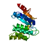
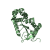
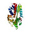
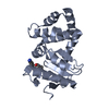
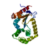
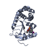
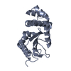
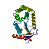
- PDB-3l9s: Crystal Structure of Salmonella enterica serovar Typhimurium DsbA -
+ Open data

- Basic information
| Entry | Database: PDB / ID: 3l9s | ||||||
|---|---|---|---|---|---|---|---|
| Title | Crystal Structure of Salmonella enterica serovar Typhimurium DsbA | ||||||
 Components Components | Thiol:disulfide interchange protein | ||||||
 Keywords Keywords | OXIDOREDUCTASE / thioredoxin-fold / DsbA / thiol-disulfide oxidoreductase / Disulfide bond / Redox-active center | ||||||
| Function / homology |  Function and homology information Function and homology informationprotein-disulfide reductase activity / cell redox homeostasis / periplasmic space Similarity search - Function | ||||||
| Biological species |  Salmonella enterica subsp. enterica serovar Typhimurium (bacteria) Salmonella enterica subsp. enterica serovar Typhimurium (bacteria) | ||||||
| Method |  X-RAY DIFFRACTION / SYNCHROTRON / MOLECULAR REPLACEMENT / molecular replacement / Resolution: 1.58 Å X-RAY DIFFRACTION / SYNCHROTRON / MOLECULAR REPLACEMENT / molecular replacement / Resolution: 1.58 Å | ||||||
 Authors Authors | Heras, B. / Jarrott, R. / Shouldice, S.R. / Guncar, G. | ||||||
 Citation Citation | Journal: J.Biol.Chem. / Year: 2010 Title: Structural and functional characterization of three DsbA paralogues from Salmonella enterica serovar typhimurium Authors: Heras, B. / Totsika, M. / Jarrott, R. / Shouldice, S.R. / Guncar, G. / Achard, M.E.S. / Wells, T.J. / Argente, M.P. / McEwan, A.G. / Schembri, M.A. | ||||||
| History |
|
- Structure visualization
| Structure viewer | Molecule: MolmilJmol/JSmol |
|---|
- Downloads & links
-Download
| PDBx/mmCIF format | 3l9s.cif.gz | 94 KB | Display | PDBx/mmCIF format |
|---|---|---|---|---|
| PDB format | pdb3l9s.ent.gz | 70.2 KB | Display | PDB format |
| PDBx/mmJSON format | 3l9s.json.gz | Tree view | PDBx/mmJSON format | |
| Others |  Other downloads Other downloads |
-Validation report
| Summary document | 3l9s_validation.pdf.gz | 415.3 KB | Display | wwPDB validaton report |
|---|---|---|---|---|
| Full document | 3l9s_full_validation.pdf.gz | 416.4 KB | Display | |
| Data in XML | 3l9s_validation.xml.gz | 11.6 KB | Display | |
| Data in CIF | 3l9s_validation.cif.gz | 17.2 KB | Display | |
| Arichive directory | https://data.pdbj.org/pub/pdb/validation_reports/l9/3l9sftp://data.pdbj.org/pub/pdb/validation_reports/l9/3l9s | HTTPS FTP |
-Related structure data
| Related structure data |  3l9uC  3l9vC  1fvkS S: Starting model for refinement C: citing same article ( |
|---|---|
| Similar structure data |


-Links
 PDBj
PDBj

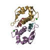
- Assembly
| Deposited unit | 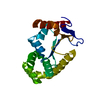
| ||||||||
|---|---|---|---|---|---|---|---|---|---|
| 1 |
| ||||||||
| Unit cell |
|
-Components
| #1: Protein | Mass: 21215.115 Da / Num. of mol.: 1 / Fragment: UNP residues 19-207 Source method: isolated from a genetically manipulated source Source: (gene. exp.) Salmonella enterica subsp. enterica serovar Typhimurium (bacteria)Strain: SL1344 / Gene: dsbA, STM3997 / Plasmid: pETLIC / Production host: References: UniProt: E1WE53, UniProt: P0A2H9*PLUS, protein disulfide-isomerase |
|---|---|
| #2: Water | ChemComp-HOH /  Mass: 18.015 Da / Num. of mol.: 259 / Source method: isolated from a natural source / Formula: H2O Mass: 18.015 Da / Num. of mol.: 259 / Source method: isolated from a natural source / Formula: H2O |
-Experimental details
-Experiment
| Experiment | Method: X-RAY DIFFRACTION / Number of used crystals: 1 |
|---|
- Sample preparation
| Crystal | Density Matthews: 2.29 Å3/Da / Density % sol: 46.38 % |
|---|---|
| Crystal grow | Temperature: 293 K / Method: vapor diffusion, hanging drop / pH: 7.5 Details: 1M ammonium sulfate, 1% (w/v) polyethylene glycol 3350, 100mM 2,2-bis(hydroxymethyl)-2,2',2"-nitrilotriethanol, pH 7.5, VAPOR DIFFUSION, HANGING DROP, temperature 293K |
-Data collection
| Diffraction | Mean temperature: 100 K |
|---|---|
| Diffraction source | Source: SYNCHROTRON / Site: Australian Synchrotron  / Beamline: MX1 / Wavelength: 0.956667 Å / Beamline: MX1 / Wavelength: 0.956667 Å |
| Detector | Type: ADSC QUANTUM 210r / Detector: CCD / Date: Aug 15, 2008 / Details: mirrors |
| Radiation | Monochromator: Double Si with sagittaly bent second crystal / Protocol: SINGLE WAVELENGTH / Monochromatic (M) / Laue (L): M / Scattering type: x-ray |
| Radiation wavelength | Wavelength: 0.956667 Å / Relative weight: 1 |
| Reflection | Resolution: 1.58→50 Å / Num. all: 25131 / Num. obs: 25131 / % possible obs: 95.3 % / Observed criterion σ(F): 0 / Observed criterion σ(I): 0 / Redundancy: 7.4 % / Rmerge(I) obs: 0.082 / Χ2: 1.064 / Net I/σ(I): 18 |
| Reflection shell | Resolution: 1.58→1.61 Å / Redundancy: 6.8 % / Rmerge(I) obs: 0.188 / Num. unique all: 1176 / Χ2: 1.09 / % possible all: 87.4 |
-Phasing
| Phasing | Method: molecular replacement | |||||||||
|---|---|---|---|---|---|---|---|---|---|---|
| Phasing MR | Model details: Phaser MODE: MR_AUTO
|
- Processing
| Software |
| ||||||||||||||||||||||||||||||||||||||||||||||||||||||||||||||||||||||||||||||||
|---|---|---|---|---|---|---|---|---|---|---|---|---|---|---|---|---|---|---|---|---|---|---|---|---|---|---|---|---|---|---|---|---|---|---|---|---|---|---|---|---|---|---|---|---|---|---|---|---|---|---|---|---|---|---|---|---|---|---|---|---|---|---|---|---|---|---|---|---|---|---|---|---|---|---|---|---|---|---|---|---|---|
| Refinement | Method to determine structure: MOLECULAR REPLACEMENT Starting model: PDB ENTRY 1FVK Resolution: 1.58→21.058 Å / Occupancy max: 1 / Occupancy min: 0.22 / FOM work R set: 0.888 / SU ML: 0.19 / σ(F): 1.91 / Stereochemistry target values: ML
| ||||||||||||||||||||||||||||||||||||||||||||||||||||||||||||||||||||||||||||||||
| Solvent computation | Shrinkage radii: 0.9 Å / VDW probe radii: 1.11 Å / Solvent model: FLAT BULK SOLVENT MODEL / Bsol: 60.27 Å2 / ksol: 0.409 e/Å3 | ||||||||||||||||||||||||||||||||||||||||||||||||||||||||||||||||||||||||||||||||
| Displacement parameters | Biso max: 53.6 Å2 / Biso mean: 16.954 Å2 / Biso min: 6.17 Å2
| ||||||||||||||||||||||||||||||||||||||||||||||||||||||||||||||||||||||||||||||||
| Refinement step | Cycle: LAST / Resolution: 1.58→21.058 Å
| ||||||||||||||||||||||||||||||||||||||||||||||||||||||||||||||||||||||||||||||||
| Refine LS restraints |
| ||||||||||||||||||||||||||||||||||||||||||||||||||||||||||||||||||||||||||||||||
| LS refinement shell | Refine-ID: X-RAY DIFFRACTION / Total num. of bins used: 9
|
