 Movie
Movie Controller

[English] 日本語
 Yorodumi
- PDB-3l6f: Structure of MHC class II molecule HLA-DR1 complexed with phospho... -
+ Open data

- Basic information
| Entry | Database: PDB / ID: 3l6f | ||||||
|---|---|---|---|---|---|---|---|
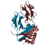
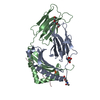
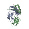
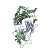
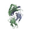
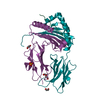
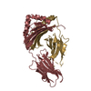
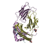
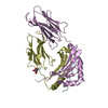
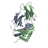
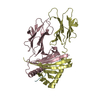
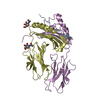
| Title | Structure of MHC class II molecule HLA-DR1 complexed with phosphopeptide MART-1 | ||||||
 Components Components |
| ||||||
 Keywords Keywords |  IMMUNE SYSTEM/PEPTIDE BINDING PROTEIN / MHC class II / HLA-DR1 / phosphopeptide / Disulfide bond / Glycoprotein / Immune response / Membrane / MHC II / Transmembrane / Endoplasmic reticulum / Golgi apparatus / IMMUNE SYSTEM-PEPTIDE BINDING PROTEIN complex IMMUNE SYSTEM/PEPTIDE BINDING PROTEIN / MHC class II / HLA-DR1 / phosphopeptide / Disulfide bond / Glycoprotein / Immune response / Membrane / MHC II / Transmembrane / Endoplasmic reticulum / Golgi apparatus / IMMUNE SYSTEM-PEPTIDE BINDING PROTEIN complex | ||||||
| Function / homology |  Function and homology information Function and homology informationregulation of interleukin-4 production / regulation of interleukin-10 production / positive regulation of T cell mediated immune response to tumor cell / myeloid dendritic cell antigen processing and presentation / antigen processing and presentation of endogenous peptide antigen via MHC class II / autolysosome membrane / regulation of T-helper cell differentiation / positive regulation of CD4-positive, CD25-positive, alpha-beta regulatory T cell differentiation / MHC class II receptor activity / positive regulation of CD4-positive, alpha-beta T cell activation ...regulation of interleukin-4 production / regulation of interleukin-10 production / positive regulation of T cell mediated immune response to tumor cell / myeloid dendritic cell antigen processing and presentation / antigen processing and presentation of endogenous peptide antigen via MHC class II / autolysosome membrane / regulation of T-helper cell differentiation / positive regulation of CD4-positive, CD25-positive, alpha-beta regulatory T cell differentiation / MHC class II receptor activity / positive regulation of CD4-positive, alpha-beta T cell activation / antigen processing and presentation of peptide or polysaccharide antigen via MHC class II / positive regulation of memory T cell differentiation / positive regulation of monocyte differentiation / CD4 receptor binding / positive regulation of kinase activity / inflammatory response to antigenic stimulus / transport vesicle membrane / intermediate filament / polysaccharide binding / T-helper 1 type immune response / Translocation of ZAP-70 to Immunological synapse / Phosphorylation of CD3 and TCR zeta chains / positive regulation of insulin secretion involved in cellular response to glucose stimulus / humoral immune response / macrophage differentiation / negative regulation of type II interferon production / Generation of second messenger molecules / immunological synapse / PD-1 signaling / epidermis development / detection of bacterium / T cell receptor binding / negative regulation of T cell proliferation / negative regulation of inflammatory response to antigenic stimulus / MHC class II antigen presentation / trans-Golgi network membrane / lumenal side of endoplasmic reticulum membrane / protein tetramerization / clathrin-coated endocytic vesicle membrane / trans-Golgi network / ER to Golgi transport vesicle membrane / structural constituent of cytoskeleton / cognition / peptide antigen assembly with MHC class II protein complex / MHC class II protein complex / positive regulation of T cell mediated cytotoxicity / peptide antigen binding / endocytic vesicle membrane / antigen processing and presentation of exogenous peptide antigen via MHC class II / Interferon gamma signaling / positive regulation of immune response / melanosome / positive regulation of T cell activation / Downstream TCR signaling / MHC class II protein complex binding / late endosome membrane / T cell receptor signaling pathway / early endosome membrane / positive regulation of canonical NF-kappaB signal transduction / adaptive immune response / positive regulation of MAPK cascade / positive regulation of viral entry into host cell / lysosome / positive regulation of ERK1 and ERK2 cascade / immune response / positive regulation of protein phosphorylation / lysosomal membrane / external side of plasma membrane / Golgi membrane / endoplasmic reticulum membrane / positive regulation of DNA-templated transcription / Golgi apparatus / cell surface / signal transduction / extracellular space / extracellular exosome / membrane / plasma membraneSimilarity search - Function | ||||||
| Biological species |  Homo sapiens (human) Homo sapiens (human) | ||||||
| Method | X-RAY DIFFRACTION / SYNCHROTRON / MOLECULAR REPLACEMENT / Resolution: 2.1 Å | ||||||
 Authors Authors | Li, Y. / Mariuzza, R.A. | ||||||
 Citation Citation | Journal: J.Mol.Biol. / Year: 2010 Title: Structural Basis for the Presentation of Tumor-Associated MHC Class II-Restricted Phosphopeptides to CD4(+) T Cells. Authors: Li, Y. / Depontieu, F.R. / Sidney, J. / Salay, T.M. / Engelhard, V.H. / Hunt, D.F. / Sette, A. / Topalian, S.L. / Mariuzza, R.A. | ||||||
| History |
|
- Structure visualization
| Structure viewer | Molecule: MolmilJmol/JSmol |
|---|
- Downloads & links
-Download
| PDBx/mmCIF format | 3l6f.cif.gz | 95.2 KB | Display | PDBx/mmCIF format |
|---|---|---|---|---|
| PDB format | pdb3l6f.ent.gz | 71.7 KB | Display | PDB format |
| PDBx/mmJSON format | 3l6f.json.gz | Tree view | PDBx/mmJSON format | |
| Others |  Other downloads Other downloads |
-Validation report
| Arichive directory | https://data.pdbj.org/pub/pdb/validation_reports/l6/3l6fftp://data.pdbj.org/pub/pdb/validation_reports/l6/3l6f | HTTPS FTP |
|---|
-Related structure data
| Related structure data |  1t5wS S: Starting model for refinement |
|---|---|
| Similar structure data |


-Links
 PDBj
PDBj


- Assembly
| Deposited unit | 
| ||||||||
|---|---|---|---|---|---|---|---|---|---|
| 1 |
| ||||||||
| Unit cell |
|
-Components
| #1: Protein | Mass: 21145.887 Da / Num. of mol.: 1 / Fragment: UNP residues 26-207 Source method: isolated from a genetically manipulated source Source: (gene. exp.) Homo sapiens (human) / Gene: HLA-DRA, HLA-DRA1 / Production host:  Escherichia coli (E. coli) / Strain (production host): BL21(DE3) / References: UniProt: P01903 Escherichia coli (E. coli) / Strain (production host): BL21(DE3) / References: UniProt: P01903 |
|---|---|
| #2: Protein | Mass: 22456.133 Da / Num. of mol.: 1 / Fragment: UNP residues 30-221 Source method: isolated from a genetically manipulated source Source: (gene. exp.) Homo sapiens (human) / Gene: HLA-DRB1 / Production host: Escherichia coli (E. coli) / References: UniProt: P04229, UniProt: P01911*PLUS |
| #3: Protein/peptide | Mass: 1665.710 Da / Num. of mol.: 1 / Fragment: UNP residues 100-114 / Source method: obtained synthetically / Source: (synth.) Homo sapiens (human) / References: UniProt: Q16655 |
| #4: Water | ChemComp-HOH / Water Mass: 18.015 Da / Num. of mol.: 196 / Source method: isolated from a natural source / Formula: H2O Mass: 18.015 Da / Num. of mol.: 196 / Source method: isolated from a natural source / Formula: H2O |
-Experimental details
-Experiment
| Experiment | Method: X-RAY DIFFRACTION / Number of used crystals: 1 |
|---|
- Sample preparation
| Crystal | Density Matthews: 2.79 Å3/Da / Density % sol: 55.87 % |
|---|---|
| Crystal grow | Temperature: 298 K / Method: evaporation / pH: 6.5 Details: 20% (w/v) PEG 8000 and 0.1 M sodium cacodylate, pH 6.5, EVAPORATION, temperature 298K |
-Data collection
| Diffraction | Mean temperature: 100 K |
|---|---|
| Diffraction source | Source: SYNCHROTRON / Site: NSLS  / Beamline: X29A / Beamline: X29A |
| Detector | Type: ADSC QUANTUM 315 / Detector: CCD / Date: Oct 30, 2009 |
| Radiation | Protocol: SINGLE WAVELENGTH / Monochromatic (M) / Laue (L): M / Scattering type: x-ray |
| Radiation wavelength | Relative weight: 1 |
| Reflection | Resolution: 2.1→43.22 Å / Num. all: 30437 / Num. obs: 30332 / % possible obs: 100 % / Redundancy: 14.2 % / Rsym value: 0.074 / Net I/σ(I): 51.3 |
| Reflection shell | Resolution: 2.1→2.18 Å / Redundancy: 14.3 % / Mean I/σ(I) obs: 5.2 / Rsym value: 0.46 / % possible all: 100 |
- Processing
| Software |
| ||||||||||||||||||||
|---|---|---|---|---|---|---|---|---|---|---|---|---|---|---|---|---|---|---|---|---|---|
| Refinement | Method to determine structure: MOLECULAR REPLACEMENT Starting model: 1T5W Resolution: 2.1→43.22 Å
| ||||||||||||||||||||
| Displacement parameters | Biso mean: 0.043 Å2
| ||||||||||||||||||||
| Refinement step | Cycle: LAST / Resolution: 2.1→43.22 Å
| ||||||||||||||||||||
| Refine LS restraints |
| ||||||||||||||||||||
| LS refinement shell | Resolution: 2.1→2.11 Å /
|
